macronucleus elongate oval to kidney-shaped, sometimes spherical or horseshoe-shaped
several spherical micronuclei scattered in the cytoplasm near the macronucleus
extrusomes rod-shaped, 6–7 µm long
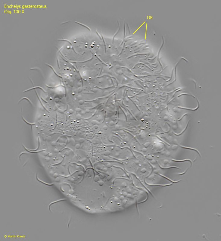
dorsal brush consisting of three rows
contractile vacuole terminal
Enchelys gasterosteus
I rarely find Enchelys gasterosteus in samples from the Simmelried. In these the samples the specimens are often found at the surface.
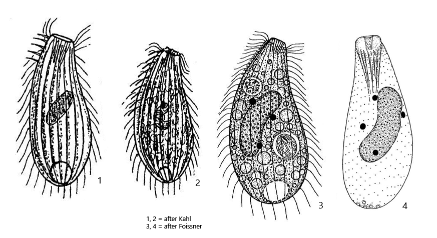
Enchelys gasterosteus was first described by Kahl (1926). Later Foissner (1984) published a redescription. In this redescription he suggests that other species were included in Kahl’s description. Thus, Kahl drew his specimens with long cilia of the dorsal brush (s. drawing 1, above). He also describes the mouth opening to be a cleft and also draws his specimens without an oral bulge (s. drawings 1 and 2, above).
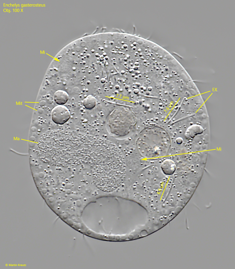
Enchelys gasterosteus is comparatively small with less than 100 µm. In my polulation the specimens were only 50–70 µm long. A typical feature of Enchelys gasterosteus are the micronuclei scattered in the plasma, which are grouped around the macronucleus. In my specimens, these micronuclei were very difficult to see in DIC because they had a refractive index similar to that of the cytoplasm (s. figs. 3 c and 4). The extrusomes in my specimens were straight as well as slightly curved rods and 6.8–7.7 µm long. Thus they correspond to the data of Foissner.
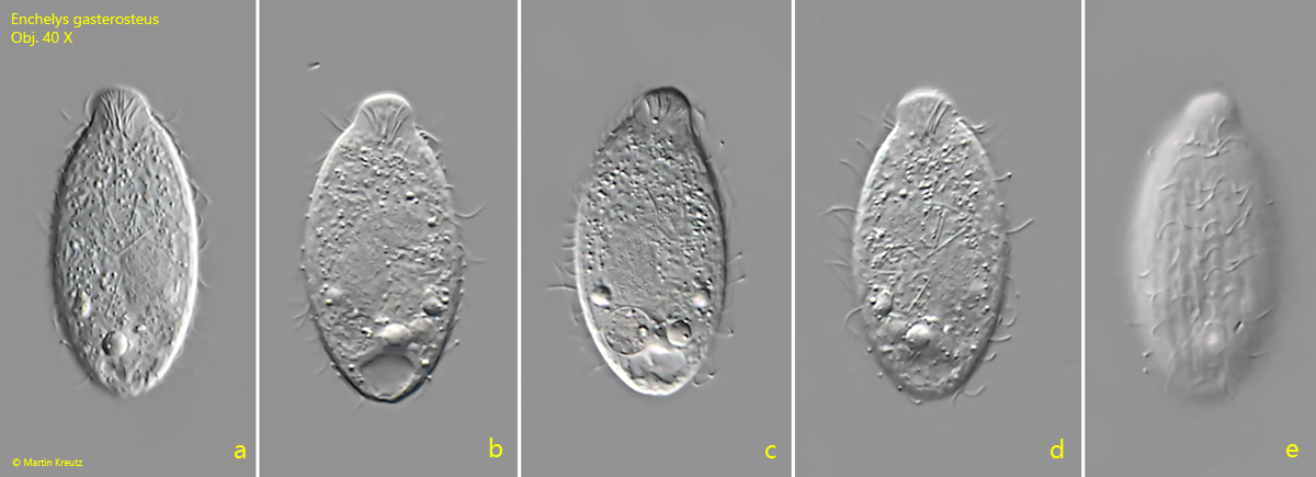
Fig. 1 a-e:Enchelys gasterosteus. L = 50 µm. A freely swimming specimen. Obj. 40 X.
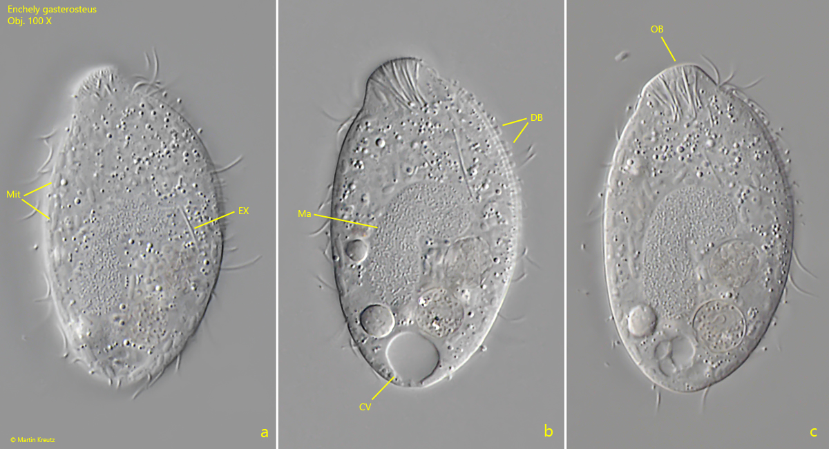
Fig. 2 a-c:Enchelys gasterosteus. L = 50 µm. Three focal planes of the slightly squashed specimen shown in fig. 1 a-e. CV = contractile vacuole, DB = dorsal brush, EX = extrusomes, Ma = macronucleus, Mit = mitochondria, OB = oral bulge. Obj. 100 X
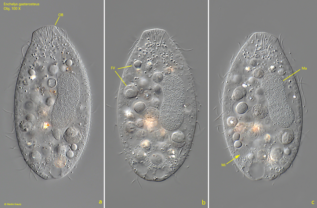
Fig. 3 a-c:Enchelys gasterosteus. L = 64 µm. Three focal planes of the slightly squashed second specimen. FV = food vacuoles, Ma = macronucleus, Mi = micronucleus, OB = oral bulge. Obj. 100 X
Fig. 4:Enchelys gasterosteus. The strongly squashed specimen shown in fig. 1 a-e. Note the micronuclei which are hard to recognize. The extrusomes (EX) of this specimen are rod-shaped and 6.9–7.7 µm long. Some of them are slightly curved. Ma = macronucleus, Mit = mitochondria. Obj. 100 X
Fig. 5:Enchelys gasterosteus. Part of the dorsal brush (DB) in a strongly squashed specimen. Obj. 100 X