cytopharynx with a tube-shaped ingestion apparatus (siphon)
siphon reaches almost the posterior end
nucleus lateral, at about mid-body
one lateral contractile vacuole in anterior third
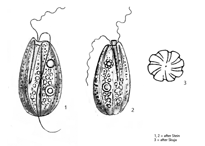
Entosiphon sulcatum
Entosiphon sulcatum is one of the most common colorless, eugenid flagellates. It occurs in almost all of my sampling sites. The species is easily identifiable by the tubular siphon and the distinct longitudinal grooves of the pellicle. I find Entosiphon sulcatum particularly often in floating detritus flakes in old samples. Entosiphon sulcatum also likes to settle on the floating coverslip.
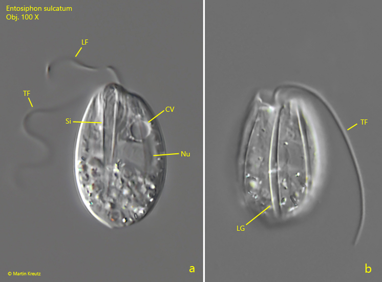
Fig. 1 a-b:Entosiphon sulcatum. L = 24 µm. Two focal planes of a slightly squashed specimen. Note the tube-shaped siphon (Si) and the longitudinal grooves (Lg) of the pellicle. CV = contractile vacuole, LF = leading flagellum, Nu = nucleus, TF = trailing flagellum. Obj. 100 X.
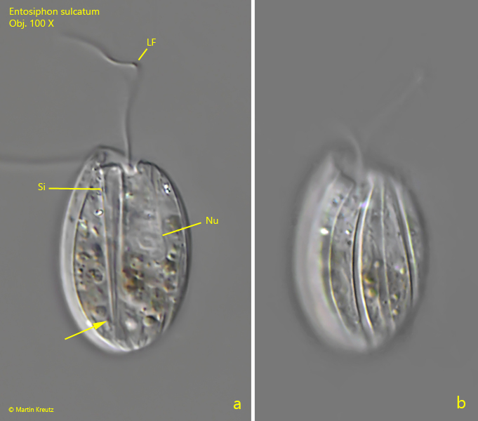
Fig. 2 a-b:Entosiphon sulcatum. L = 27 µm. Two focal planes of a second specimen. Note that the siphon (Si) is reaches almost down to the posterior end (arrow). LF = leading flagellum, Nu = nucleus. Obj. 100 X.
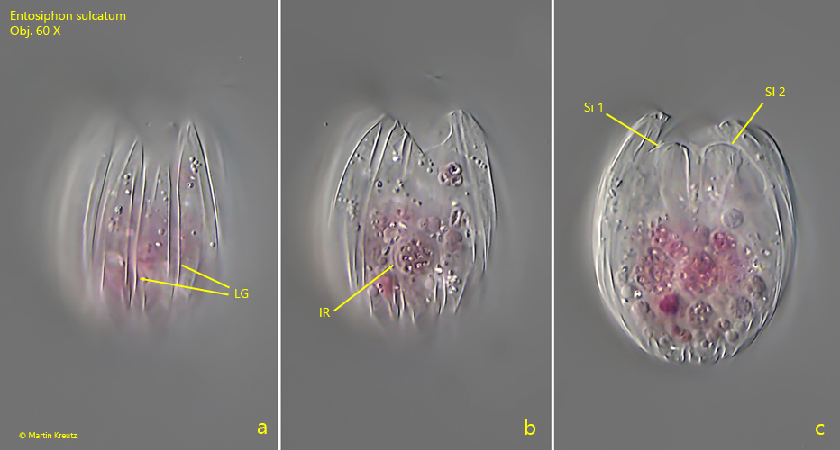
Fig. 3 a-c:Entosiphon sulcatum. L = 28 µm. Three focal planes a slightly squashed specimen in the process of cell division. Note the doubled siphons (Si 1, Si 2). The specimen appears pink due to ingested rhodobacteria (IR). LG = longitudinal grooves of the pellicle. Obj. 60 X.