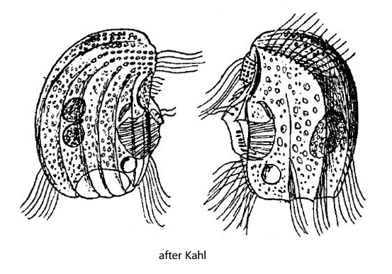
Kahl decribed the odontostomatid ciliate Epalxella striata to be frequent in occurrence. However, in my sampling area Simmelried I have found this species only very rarely. In 1995 I found a specimen in the uppermost mud layer. At that time I could document the right side (s. fig. 1a-b). It was not until 25 years later that I found a second specimen in 2020 to photograph the left side (s. fig. 2 a-f) and an ventral view (s. fig. 3 a-c). Since then I have not found further specimens.
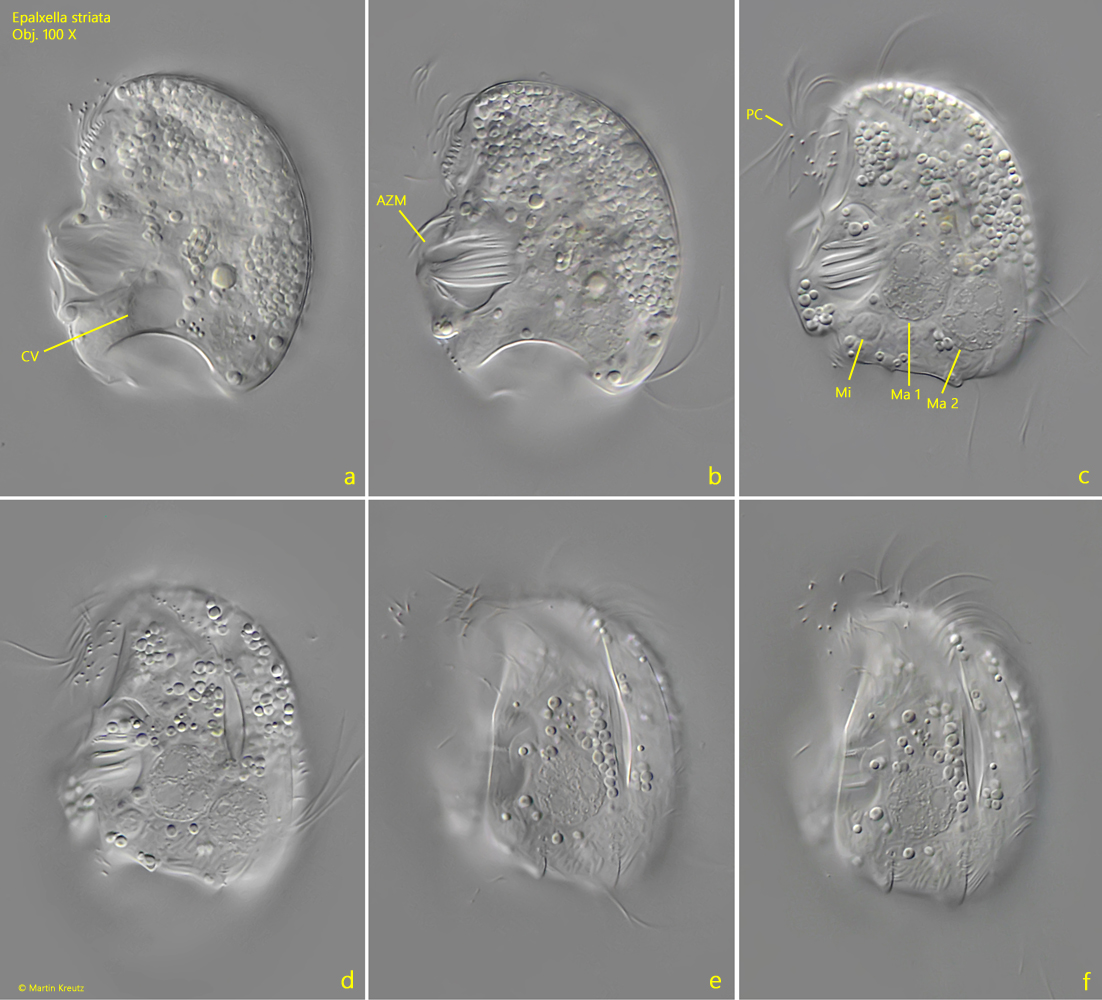
A characteristic feature of Epalxella striata are the protrusions of the pellicle on the left side, where the longitudinal ridges meet the posterior margin. This gives the impression that the pellicle is folded at the posterior margin (s. fig. 2 e and 2 f).
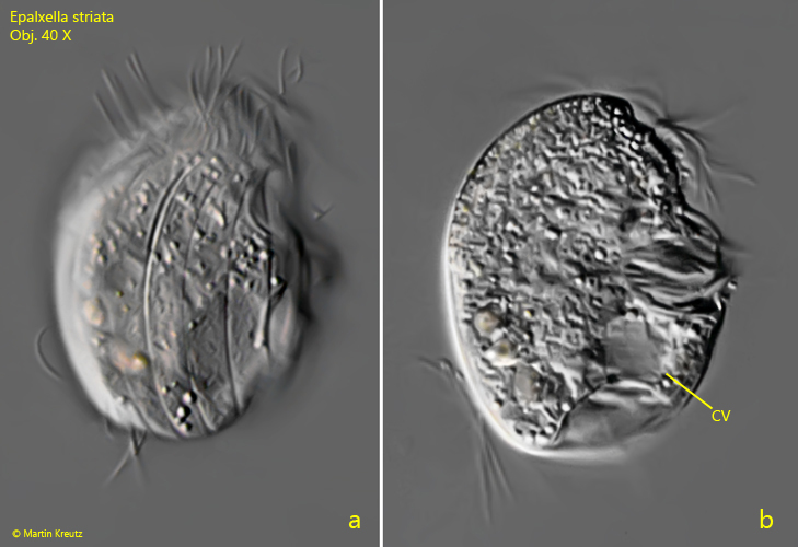
Fig. 1 a-b:Epalxella striata. L = 28 µm. Lateral view from the right side. CV = contractile vacuole. Obj. 40 X.
Fig. 2 a-f:Epalxella striata. L = 42 µm. Different focal planes from left. AZM = adoral zone of membranelles, Ma 1 + Ma 2= macronuclei, Mi = micronucleus, PC = perizonal cilia row. Obj. 100 X.
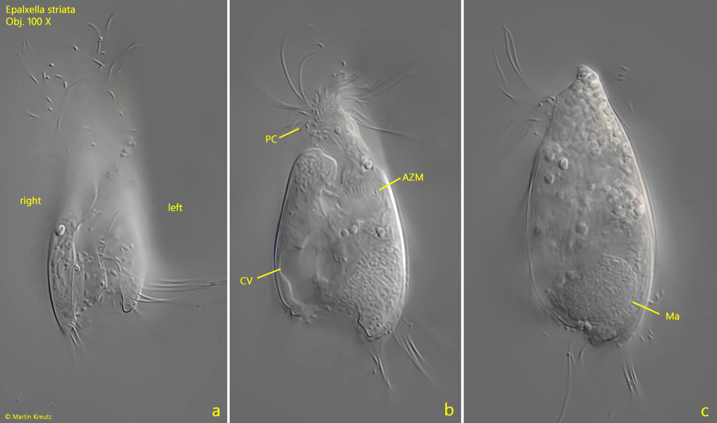
Fig. 3 a-c:Epalxella striata. L = 50 µm. Different focal planes from ventral of a freely swimming specimen. AZM = adoral zone of membranelles, CV = contractile vacuole, Ma = macronucleus, PC = perizonal cilia row. Obj. 100 X.