cells spindle-shaped, anterior end obliquely truncated
cells attached with a tapered end to inner wall of lorica
cells 28–35 µm long (with tapered ends)
lorica 70–90 µm long, cylindrically, straight or slightly curved
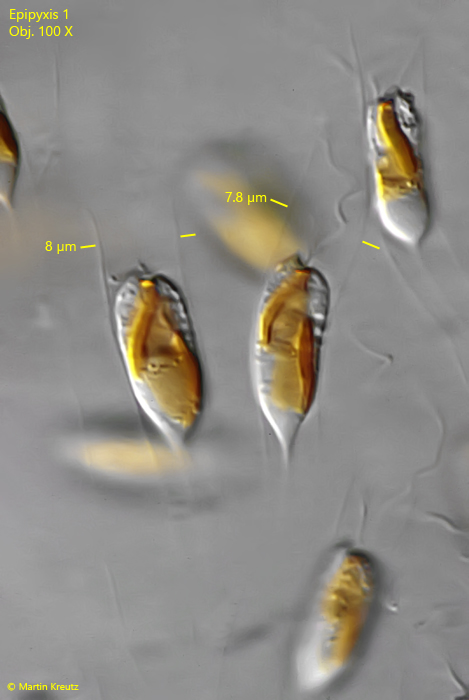
aperture of lorica slightly not widened, diamater 6.5–8 µm
loricae in parallel bundles
one chloroplast
one contractile vacuole in anterior half
one eyespot
two flagella of different length
colonies trumpet-shaped or conical
No drawings from previous autors available.
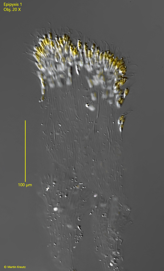
In April 1995, I found several colonies of a chrysophyte in samples from Ulmisried. The monads were located in tubular loricae, which were grouped together in almost parallel bundles (s. fig. 1). The colonies reached a length of up to 800 µm. In November 1999, I also found the same species in Simmelried. After 1999, I have not been able to find this species again.
I found the colonies both freely swimming and attached to plant stems. It is possible that the freely swimming colonies detached from the substrate during the transport of the samples. Although the loricae appear smooth and without a scale structure, I believe that the genus Epipyxis is present here, as the scale structure typical for Epipyxis loricae is not or only barely visible under the light microscope.
The loricae of this Epipyxis species are 70–90 µm long (difficult to discern) and have a diameter of 6.5–8 µm. The apertures of the loricae is not or only very slightly widened. The most striking feature is the nearly parallel arrangement of the loricae in bundles. The colonies are neither branched nor bushy. In addition, the bundled loricae remain together over several cell generations, which can cause the colonies to become very thick and long.
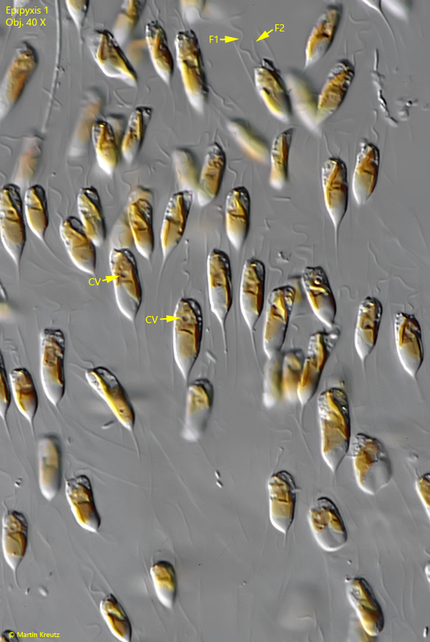
The monads have a length of 28–35 µm with their tapered end and sit near the end of the loricae. They possess only one chloroplast and a contractile vacuole in the anterior half, often in the anterior third (s. figs. 4 and 5).
Among the described Epipyxis species, I could not find any species with these characteristics. Especially the parallel arrangement of the loricae within the colonies seems unique. Therefore, I consider it possible that this represents a previously undescribed species Epipyxis nov. spec.
Fig. 1:Epipyxis 1. L = 390 µm (of colony). A colony of two parallel bundles of loricae. Obj. 20 X.
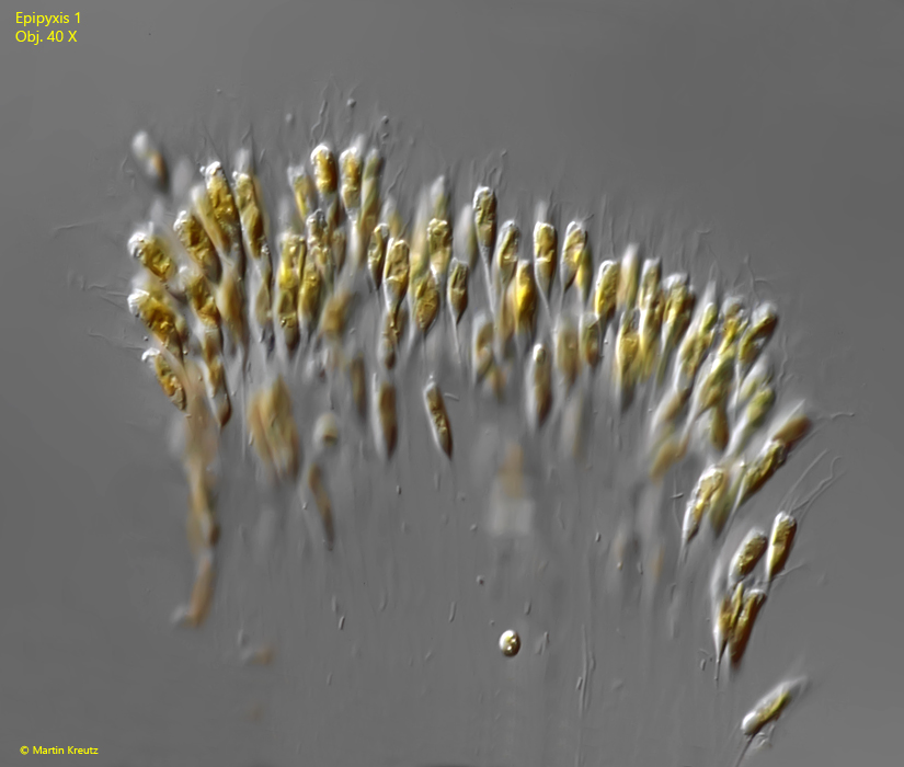
Fig. 2:Epipyxis 1. The cells located near the end of the loricae and are attached with the tapered end to the inner wall. of them. Obj. 40 X.
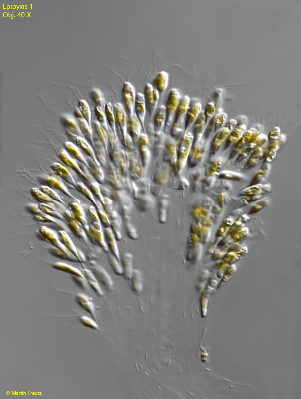
Fig. 3:Epipyxis 1. The cells of a second colony. Obj. 40 X.
Fig. 4:Epipyxis 1. The spindle-shaped cells have two flagella (F1, F2) of different length and a single contractile vacuole (CV) in the anterior half. Obj. 40 X.
Fig. 5:Epipyxis 1. The loricae are tube-shaped with a diameter of 6.5-8 µm. The aperture of the loricae is not or only very slightly widened. In the cells only one chloroplast is visible. Obj. 100 X.