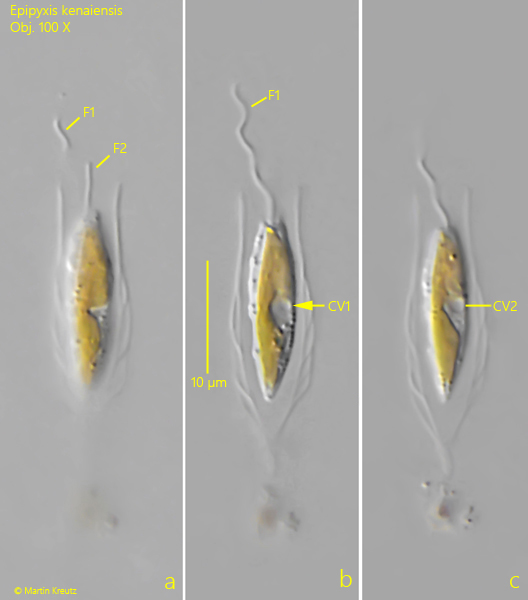
On the wall of a vessel containing an old sample from the Schwemm Moor, I found a single specimen of Epipyxis kenaiensis in July 2025. Since the species lives solitarily, it can easily be overlooked among detritus and algae in fresh samples.
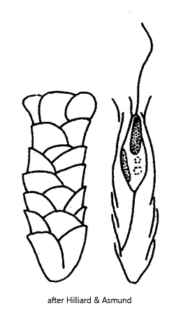
The description of Epistylis kenaiensis by Hillilard and Asmund (1963) is very brief. An important characteristic is the structure of the lorica, which is made up of round scales; these can only be seen in an optical section of the lorica in lateral view (s. fig. 1 b). The round scales are very delicate and difficult to contrast in DIC. Another important characteristic is the shape and width of the aperture. In my specimen, the lorica aperture was slightly widened and had a diameter of 4.8 µm. In the similar species Epipyxis borealis, the diameter of the aperture is a maximum of 3.5 µm.
The cell is spindle-shaped and has 2 contractile vacuoles near the middle of the body. In my specimen, they were superimposed from the viewing angle, but still visible (s. figs. 1 b and 1 c). A small eyespot sits apically on the upper chromatophore. There are two flagella of different lengths (s. fig. 1 a).
Fig. 1 a-c:Epipyxis kenaiensis. L = 25 µm (of lorica). Three focal planes of a spindle-shaped specimen. Note the the two (superimposed) contractile vacuoles in mid-body. The aperture of the lorica is 4.8 µm wide. F1, F2 = flagella of unequal length. Obj. 40 X.