So far, I have found
Epipyxis utriculus var.
acuta only very rarely and exclusively in the
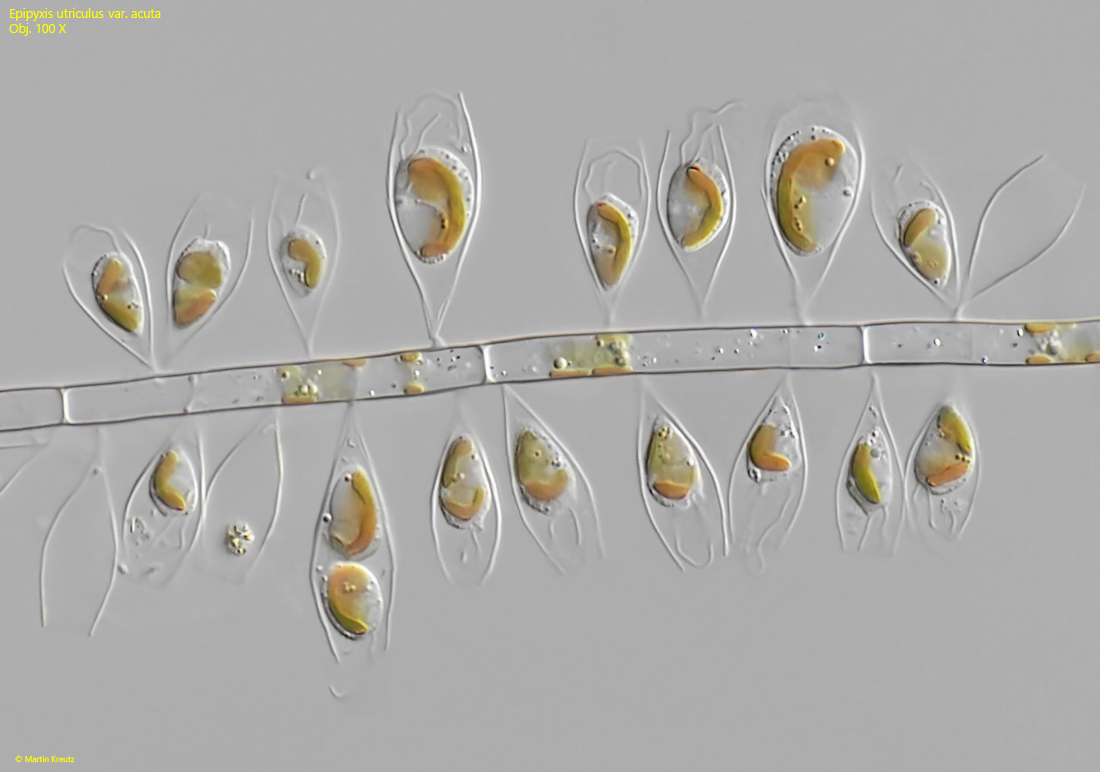
Simmelried. There, I mostly find the specimens on algal filaments, which are often densely colonized (s. fig. 1).
Epipyxis utriculus var.
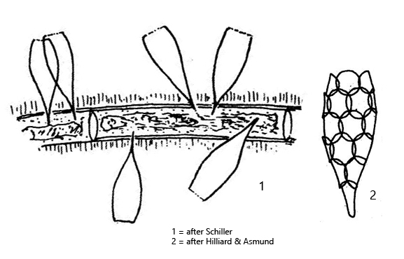
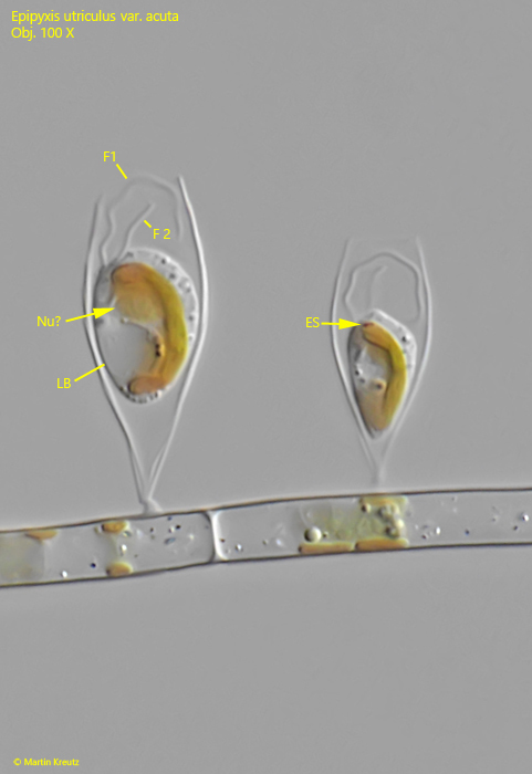
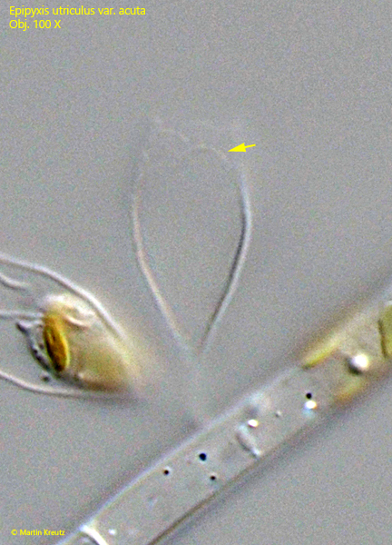
acuta can be recognized by the typical shape of its lorica. It somewhat resembles an ice cream cone, with the front third slightly narrowed. The evenly tapering rear part of the shell ends in an adhesive disc, with which the specimens attach themselves to the substrate. The lorica is assembled of very delicate, round scales that can only be seen with staining or in phase contrast (s. drawing 2 above). In DIC, these scales are difficult to visualize (s. fig. 4).
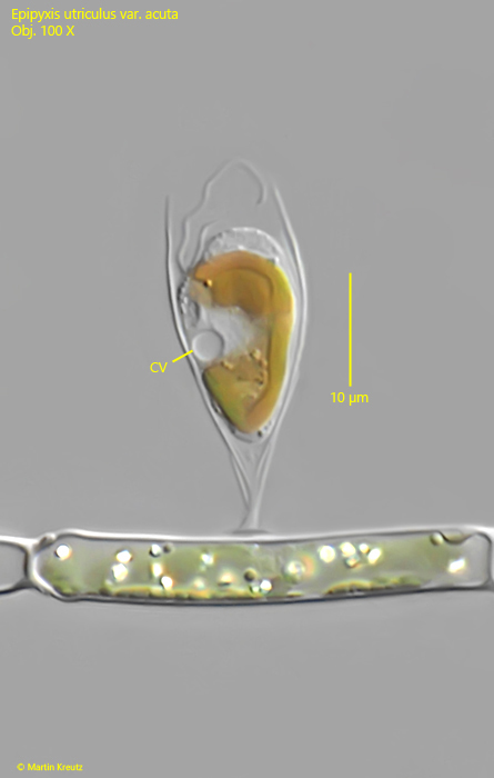
The cells are spindle-shaped, with the hyaline posterior end being difficult to see because it tapers almost thread-like. The cell is attached to the bottom of the lorica. There is only one chloroplast, topped by a small eyespot (s. fig. 3). The cells have two flagella of different lengths, and the contractile vacuole is located near the middle of the body (s. fig. 2).