fragmented macronucleus of about 100 small nodules
about 10–20 micronuclei
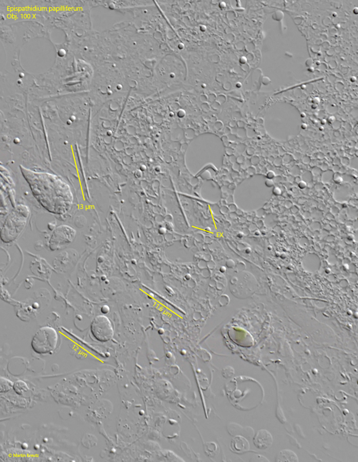
extrusomes straight, rod-shaped, 10–12 µm long
contractile vacuole terminal
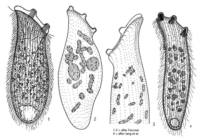
Epispathidium papilliferum
I found Epispathidium papilliferum in moss taken from a tree trunk. The moss was moistened with water from the Simmelried. It took 2 weeks for a population of Epispathidium papilliferum to develop. Besides Epispathidium papilliferum, I was only able to detect large numbers of Halteria grandinella, Colurella uncinata, and Cyclidium glaucoma in the moss sample.
Originally, Epispathidium papilliferum was described as Spathidium papilliferum by Kahl in 1930. In 1984, the species was then transferred to the genus Epispathidium by Foissner.
The specimens of my population were 150–200 µm long. A characteristic feature of Epispathidium papilliferum is the 3 papillae of the oral bulge. However, in my population, the dorsal papilla was only weakly developed (s. fig. 3 a). This observation was also made by Kahl and Foissner, who also found specimens with only two papillae. There is evidence that the number and shape of the papillae depend on the food supply. Kahl reported that starving forms of Epispathidium papilliferum even have finger-shaped, elongated papillae.
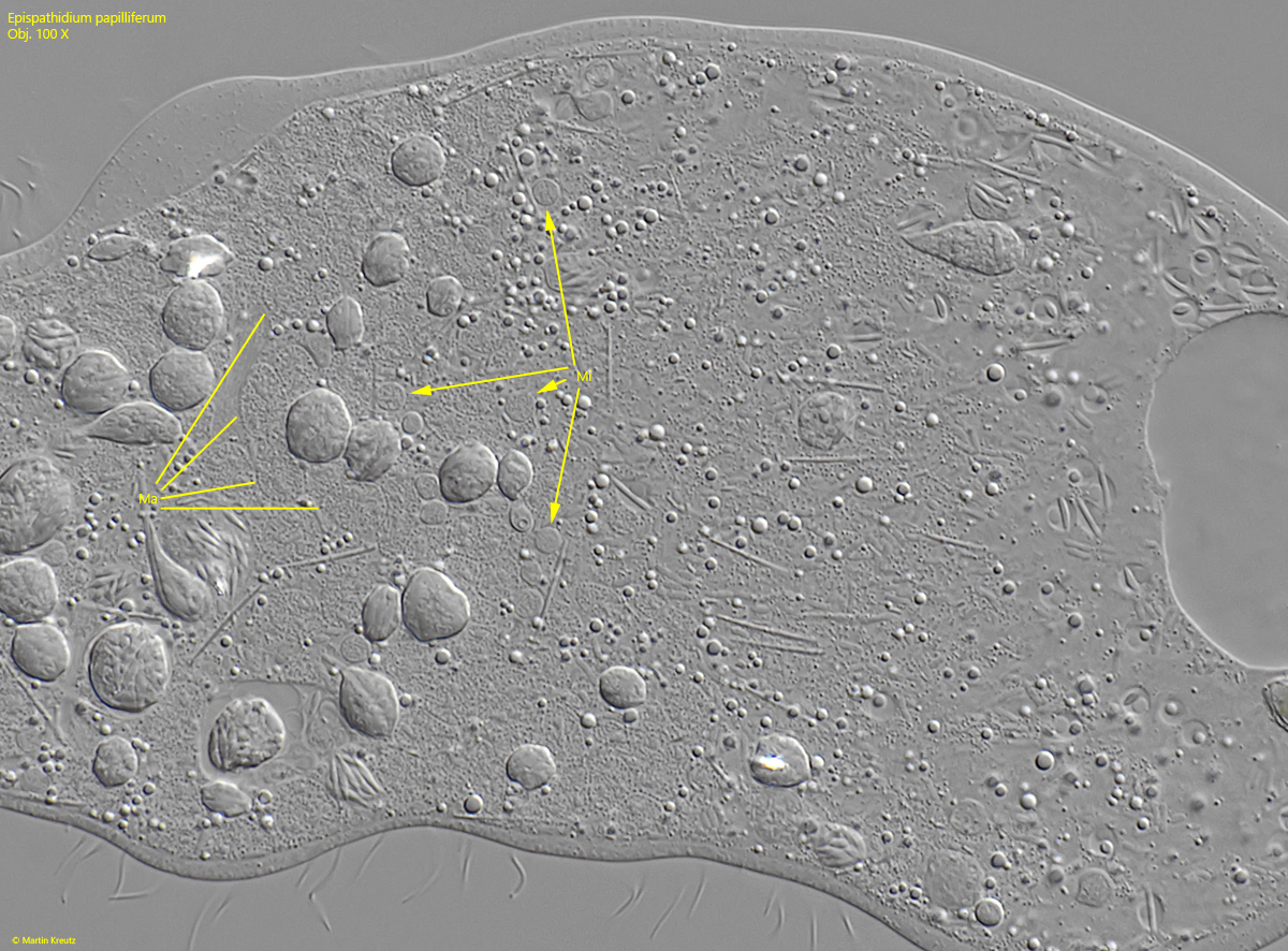
The nodules of the macronucleus were very numerous and small in my specimens. They were also not easy to recognize in heavily compressed specimens (s. fig. 4). However, the small, spherical micronuclei were always clearly visible (s. fig. 4). The straight, rod-shaped extrusomes in my population were 11–13 µm long and thus corresponded exactly to the descriptions by Jang et al. (2017), Kahl and Foissner (s. fig. 5).
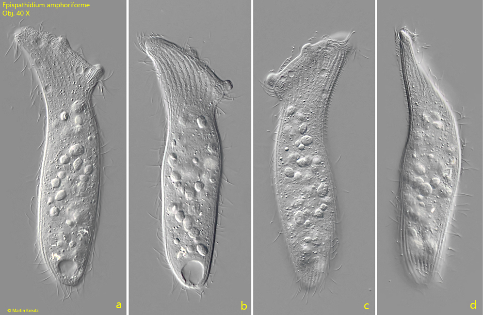
Fig. 1 a-d:Epispathidium papilliferum. L = 175 µm. A freely swimming specimen from right (a, b), left (c) and from dorsal (d). Obj. 40 X.
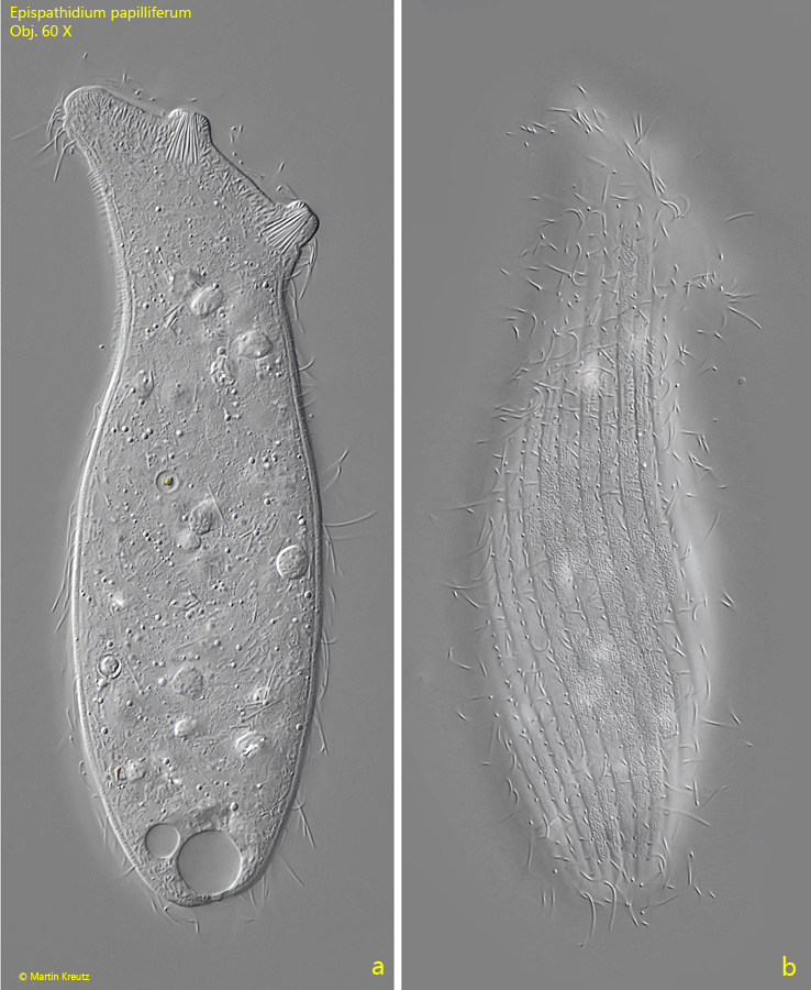
Fig. 2 a-b:Epispathidium papilliferum. L = 195 µm. Two focal planes of a second, slightly squashed specimen from right. Obj. 60 X.
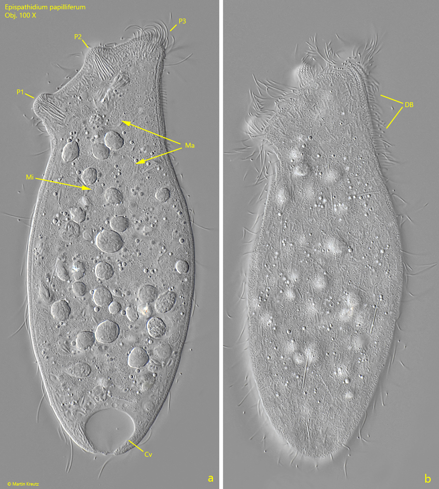
Fig. 3 a-b:Epispathidium papilliferum. L = 152 µm. A squashed third specimen from left. From the three papillae (P1–P3) the dorsal papilla P3 is reduced. The small nodules of the macronucleus (Ma) are visible as well as a micronucleus. CV = contractile vacuole, DB = dorsal brush. Obj. 100 X.
Fig. 4 a-b:Epispathidium papilliferum. In a squashed specimen some of the macronuclear nodules (Ma) are visible as well some of the micronuclei (Mi). Obj. 100 X.
Fig. 5:Epispathidium papilliferum. The extrusomes are straight, rod-shaped and have a length of 11–13 µm. Obj. 100 X.