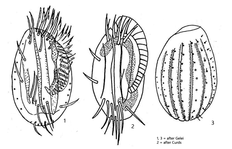
dorsally 6–7 ribs, with 8 rows of short bristles between
9 frontoventral cirri
1 (somtimes 2) marginal cirrus
5 transverse cirri
2 caudal cirri
8 (rarely 6 or 7) dorsal rows of short bristles
macronucleus C-shaped
one spherical micronucleus adjacent to macronucleus
contractile vacuole at level of transverse cirri
Euplotes affinis
Euplotes affinis is a very common hypotrichous ciliate, which is easy to overlook in fresh samples due to its small size. However, it likes to settle on the floating coverslip and is also frequently found near the surface of old samples.
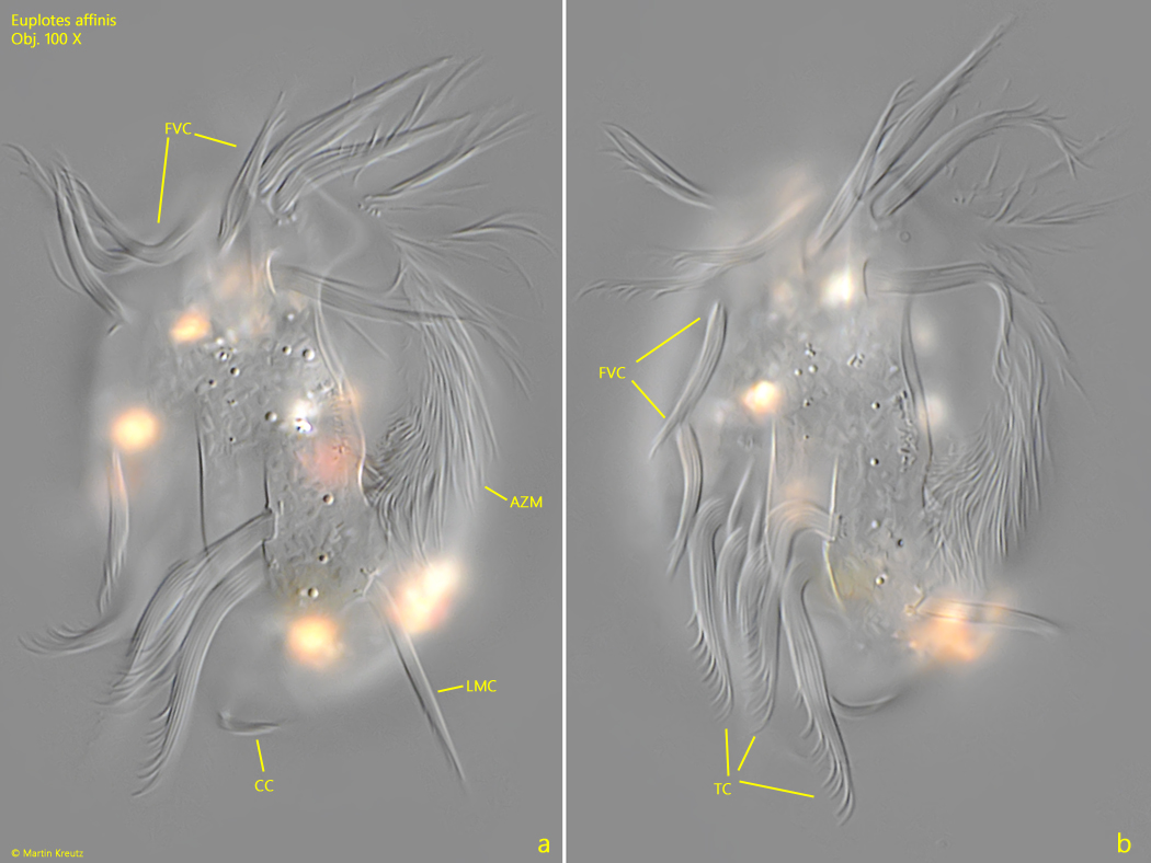
A characteristic feature is the spread-out, separate left marginal cirrus, which stands out due to the otherwise greatly reduced cilia (s. fig. 2 a). Of the total of 9 frontoventral cirri, two are clearly separated on the right margin of the body (s. fig. 2 b). Longitudinal ribs are located between the strong transverse cirri (s. fig. 2 b). The dorsal side is convexly curved and also has ribs (s. fig. 4). Between the dorsal ribs are rows of short, bristle-like cilia, which can only be seen under high magnification in squashed specimens (s. fig. 5). The macronucleus is C-shaped, which can also only be seen in squashed specimens. The micronucleus is often found at one end of the macronucleus, but is difficult to see as it can often be very hyaline (s. fig. 6).
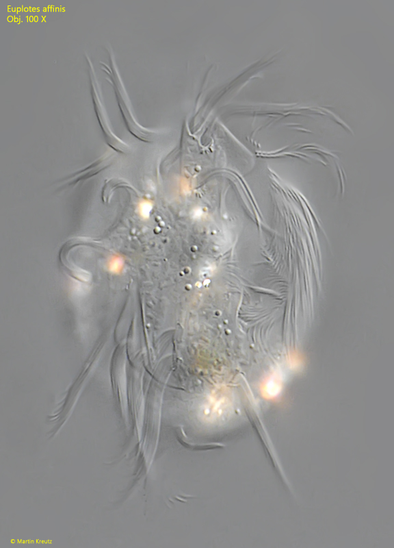
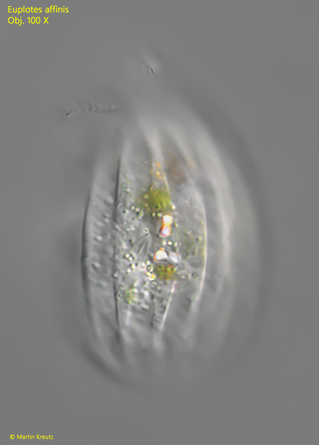
Fig. 1:Euplotes affinis. L = 55 µm. A freely swimming specimen from ventral. Obj. 100 X.
Fig. 2 a-b:Euplotes affinis. L = 55 µm. Two focal planes of a slightly squashed specimen from ventral. Note the separate left marginal cirrus (LMC), which is often spread apart in resting specimens. Two of the nine frontoventral cirri (FVC) are separated on the right margin (fig. 1 b). AZM = adoral zone of membranelles, BC = buccal cirrus, CC = caudal cirri, TC = transversal cirri. Obj. 100 X.
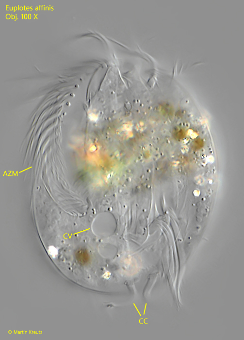
Fig. 3:Euplotes affinis. A squashed specimen from dorsal to show the position of the contractile vacuole, slightly shifted to the right side. AZM = adoral zone of membranelles, CC = caudal cilia. Obj. 100 X.
Fig. 4:Euplotes affinis. L = 58 µm. The dorsal ribs of a frely swimming specimen. Obj. 100 X.
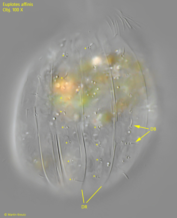
Fig. 5:Euplotes affinis. A squashed specimen from dorsal. Between the dorsal ribs (DB) rows of short bristles are located (DB). The rows are labelled with “*”. Obj. 100 X.
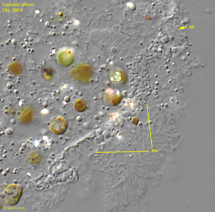
Fig. 6:Euplotes affinis. The macronucleus (Ma) in a strongly squashed specimen and the hyaline micronucleus (Mi) located at the end. Obj. 100 X.