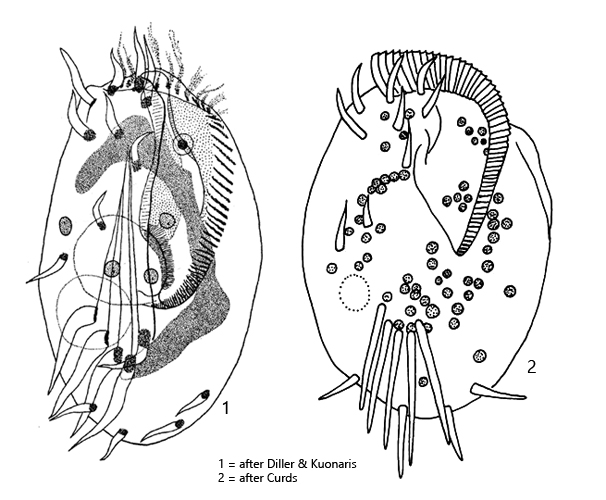
contractile vacuole on right side, posterior third
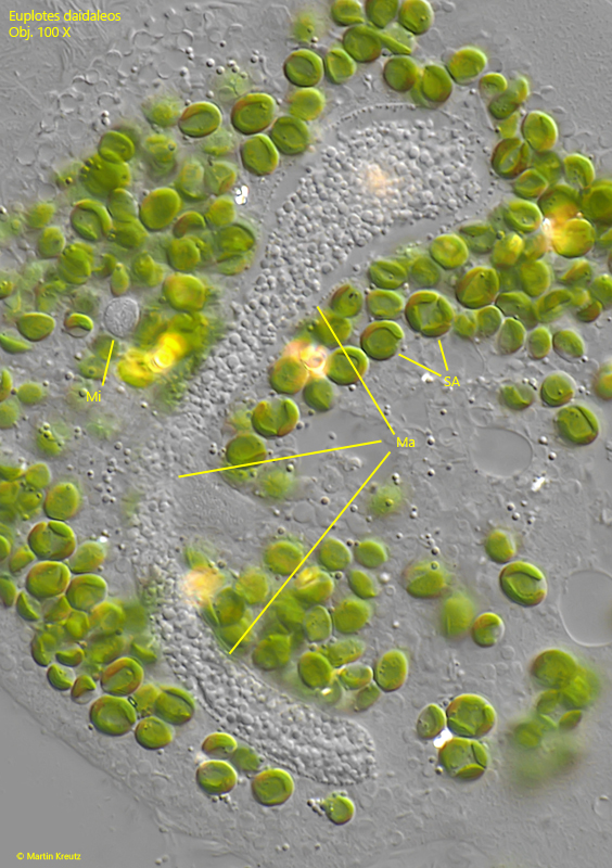
macronucleus C-shaped
micronucleus in anterior third with distance to macronucleus
6 frontal cirris
2 frontoventral cirri
1 buccal cirrus
5 transverse cirri
2 left marginal cirri
2 caudal cirri
6 ridges between transversal cirri
dorsal side with 6–7 rows of short bristles
Euplotes daidaleos
Euplotes daidaleos is a very common hypotrich ciliate in my sampling sites. I mostly find it among decaying leaves or in the top layer of mud. Unlike many other hypotrich ciliates, Euplotes daidaleos does not settle or only very rarely settles on the floating coverslip.
It was only in 1966 that Euplotes daidaleos was recognized and described as a distinct species by Diller and Kuonaris. This is somewhat surprising because Euplotes daidaleos is commonly found worldwide. The species mainly differs from the very similar species Euplotes patella and Euplotes aediculatus by the possession of symbiotic algae. The ciliation corresponds to that of Euplotes patella. Only the number of adoral membranelles is said to be higher in Euplotes daidaleos, which is difficult to verify in living specimens.
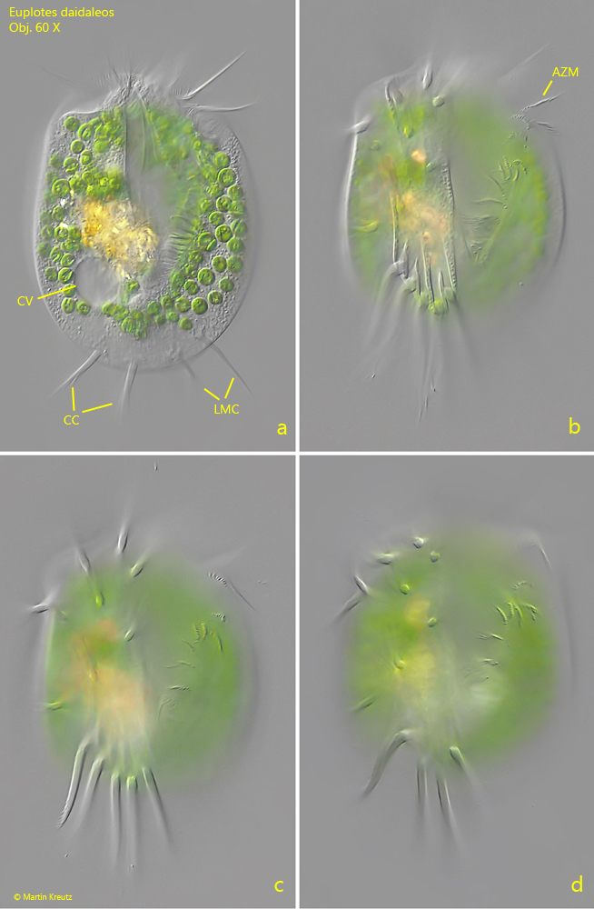
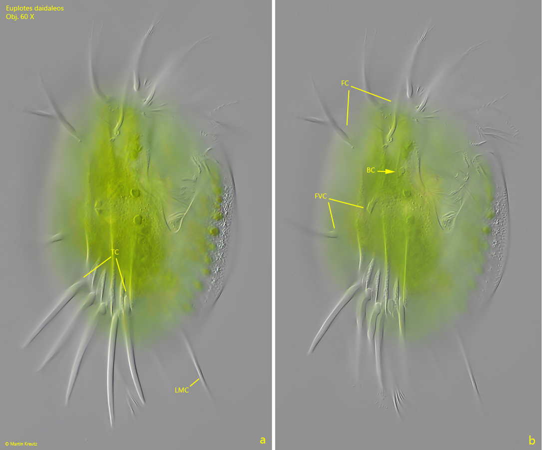
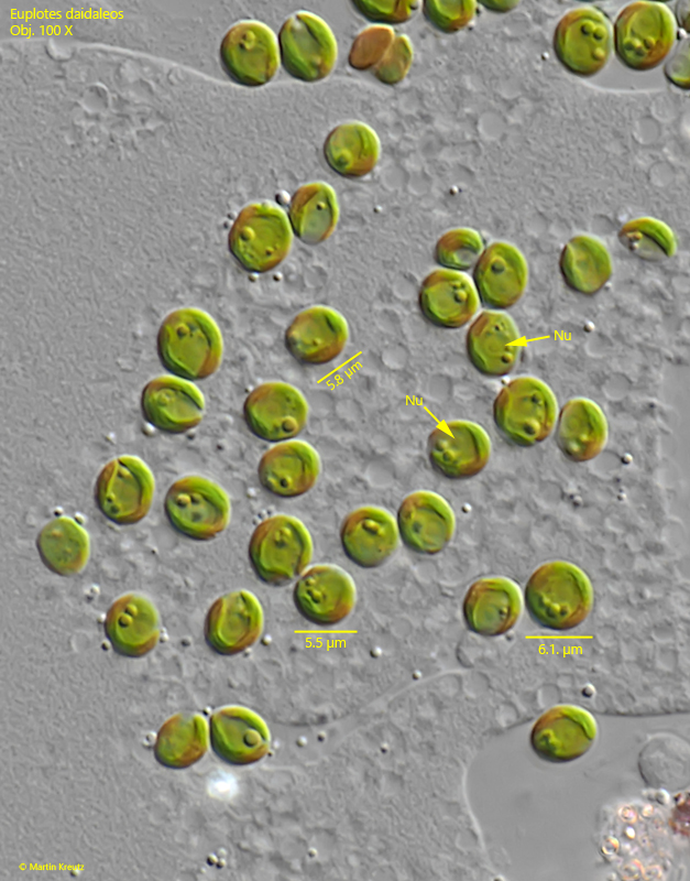
The specimens of my population were between 90–130 µm long. The body shape varied between broadly elliptical and almost rectangular with nearly parallel sides. I was able to clearly see the 6 frontal cirri, the two frontoventral cirri, and the buccal cirrus (s. fig. 2 a-b). At the posterior end, there are 2 left marginal cirri and on the right side two caudal cirri. The 5 transverse cirri clearly protrude beyond the posterior end and are usually somewhat spread apart (s. fig. 2 a-b). Additionally, the distal end of the transverse cirri is often frayed. The macronucleus is long and C-shaped. The small micronucleus is always found at some distance from the macronucleus (s. fig. 5). The symbiotic algae had a diameter of 5.5–6.1 µm and corresponded to the Chlorella type with its own cell nucleus (s. fig. 6).
Fig. 1 a-d:Euplotes daidaleos. L = 89 µm. Different focal planes of a freely swimming specimen from ventral. In the cytoplasm many symbiotic algae are scattered. The adoral zone of membranelles (AZM) reach almost the posterior third of the body. At the posterior end the 2 left marginal cirri (LMC) as well as the two caudal cirri (CC) are visible. The contractile vacuole is located on the right side. Obj. 60 X.
Fig. 2 a-b:Euplotes daidaleos. L = 128 µm. Two slightly different focal planes from ventral. There are 6 frontal cirri (FC), one buccal cirrus (BC), 2 frontoventral cirri (FVC) and 5 transverse cirri (TC) visible. One of the left marginal cirri (LMC) is also visible. Obj. 60 X.
Fig. 3:Euplotes daidaleos. L = 129 µm. A third specimen from ventral with focal plane on the adoral zone of membranelles (AZM). Obj. 60 X.
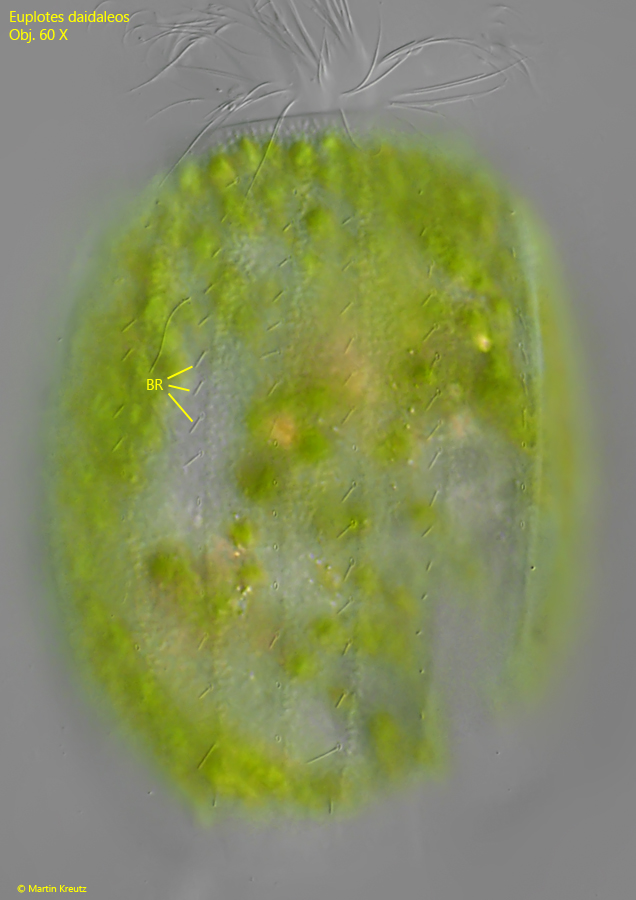
Fig. 4:Euplotes daidaleos. L = 110 µm. A slightly squashed specimen from dorsal. Note the 6 rows of short bristles (BR) between flat ribs. Obj. 60 X.
Fig. 5:Euplotes daidaleos. The C-shaped macronucleus (Ma) and the spherical micronucleus (Mi) in a squashed specimen. Obj. 100 X.
Fig. 6:Euplotes daidaleos. The symbiotic algae scattered in the cytoplasm have a diameter of 5.5–6.1 µm with a cup-shaped chloroplast. They seems to be of the Chlorella type. Each alga cell has an own nucleus (Nu). Obj. 60 X.