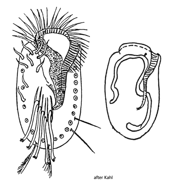
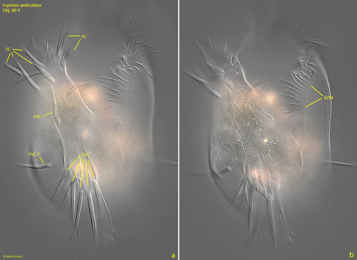
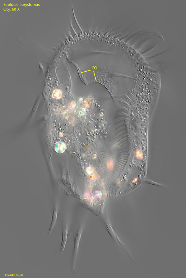
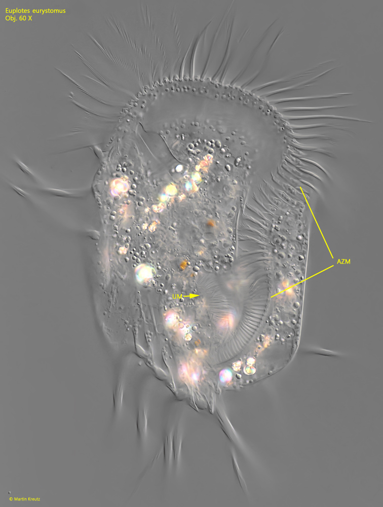
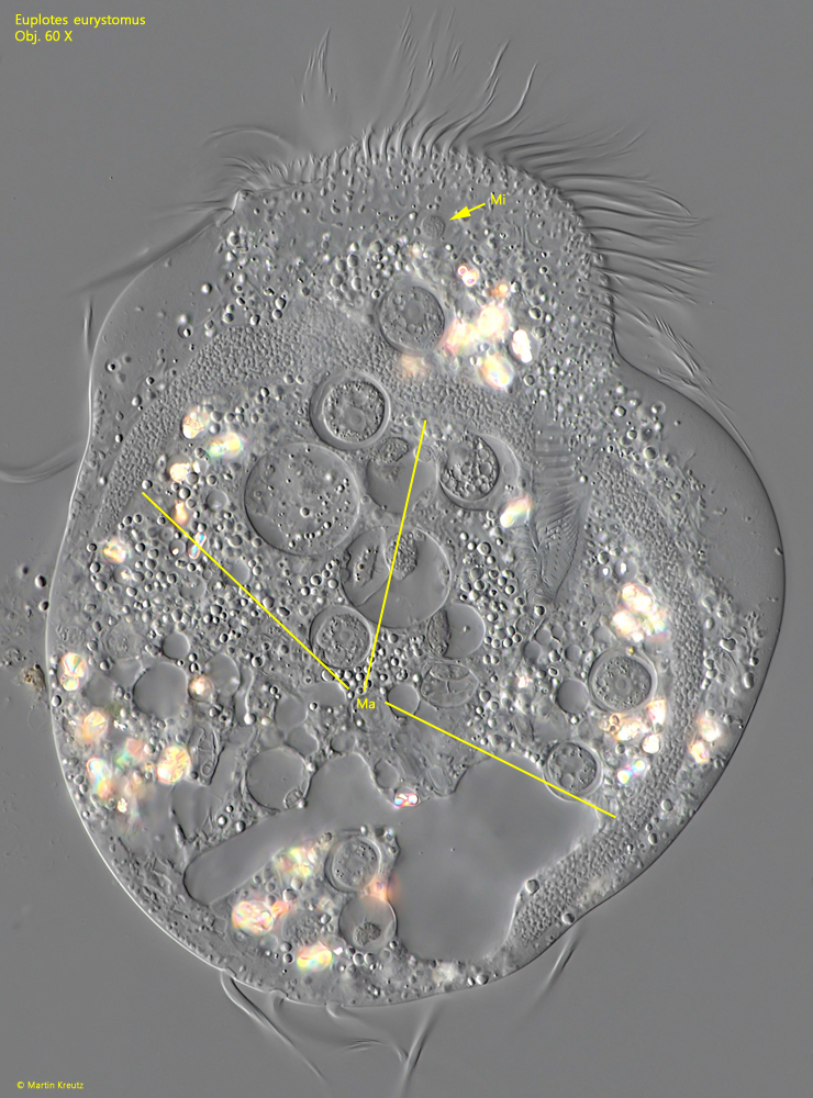
Euplotes eurystomus
Wrzesniowski, 1870
Most likely ID: n.a.
Synonym: n.a.
Sampling location: Pond of the waste disposal company Constance
Phylogenetic tree: Euplotes eurystomus
Diagnosis:
- body ellipsoid, almost parallel sided
- length 140–180 µm, width 95–135 µm
- apically a collar-shaped protrusion
- oral field large, triangular, in middle with convex fold
- large adoral zone, S-shaped, reach to posterior third
- 6 frontal cirri
- 1 buccal cirrus
- 2 frontoventral cirri
- 5 transversal cirri between 6 ridges
- 2 caudal cirri
- 2 left marginal cirri
- macronucleus C-shaped or 3-shaped
- small, spherical micronucleus in anterior third
- contractile vacuole in anterior third, right
I have only found Euplotes eurystomus once, in an old sample with decomposing water lily leaves, together with a large population of Paramecium aurelia. According to Foissner et al. (1991), Euplotes eurystoma is a rather rare species within the genus. The authors themselves did not find any specimens.
Euplotes eurystomus is notable for a very large, triangular oral field, which on the left side is bordered by a very pronounced adoral zone running in a distinct S-shape (s. fig. 5). Additionally, I noticed that the specimens often swim freely and do not walk along glass surfaces as frequently. The 5 transverse cirri are long and extend clearly beyond the posterior end (fig. 3 a-b). Furthermore, there are 2 caudal cirri at the posterior end and two marginal cirri on the left side (s. fig. 2 d). On the ventral side, a total of 9 cirri can be seen, divided into 6 frontal cirri, 1 buccal cirrus, and 2 frontoventral cirri (s. fig. 3 a). The latter are quite far apart from each other.
In the cytoplasm, I could identify the symbiotic bacteria mentioned by Foissner et al. (s. fig. 9). According to my observations, they are 8–10 µm long and about 1 µm thick. In the food vacuoles, I very frequently found phagocytized amoebae of the genus Nuclearia. Additionally, in the cytoplasm of all specimens of the population, round, strongly birefringent bodies of unknown composition were found. Some were vaguely dumbbell-shaped (s. fig. 7).
Fig. 1 a-d: Euplotes eurystomus. L = 185 µm. A freely swimming specimen with an elliptical shaped body from ventral. On the left side the two marginal cirri are visible (LMC 1, LMC 2). Note the collar-shaped protrusion of the anterior end (CO). CV = contractile vacuole. Obj. 40 X.
Fig. 2 a-d: Euplotes eurystomus. L = 150 µm. A freely swimming specimen with an broadly elliptical body from ventral. Note the two marginal cirri (LMC 1, LMC 2) and the two caudal cirri (CC). Obj. 60 X.
Fig. 3 a-b: Euplotes eurystomus. L = 194 µm. The pattern of the ventral cirri of a slightly squashed specimen. There 6 frontal cirri (FC), one buccal cirrus (BC) and two, separated frontoventral cirri (FVC 1, FVC 2). The 5 transversal cirri (TVC) arise between longitudinal ridges. AZM = adoral zone of membranelles. Obj. 60 X.
Fig. 4: Euplotes eurystomus. L = 185 µm. On the right side of the oral field a convex fold (FO) is located. Obj. 60 X.
Fig. 5: Euplotes eurystomus. L = 148 µm. The adoral zone of membranelles has an S-shape. In the posterior third the inconspicuous undulating membrane (UM) is visible. Obj. 60 X.
Fig. 6: Euplotes eurystomus. The long C-shaped macronucleus (Ma) and the small spherical micronucleus (Mi) in a squashed specimen. Obj. 60 X.
Fig. 7: Euplotes eurystomus. The macronucleus (Ma) and micronucleus (Mi) in a second, squashed specimen. Obj. 100 X.
Fig. 8: Euplotes eurystomus. The dosal side of a slightly squashed specimen. From the 10 dorsal rows of cilia 5 are visible (arrows). The dorsal cilia are short as bristles (BR) and arise from pores. Obj. 100 X.
Fig. 9: Euplotes eurystomus. The symbiotic bacteria (SB) in the cytoplasm are 8–10 µm long and slightly cureved. Obj. 100 X.