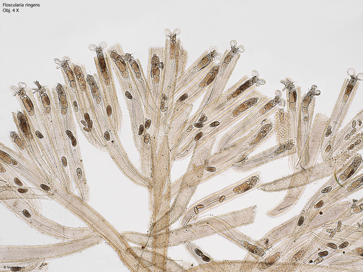
In my sampling site Simmelried, I only ever find Floscularia ringens as solitary specimens, mostly attached to leaves of Utricularia. However, in samples from the Schwemm Moor in Austria, I have also found very large, branched colonies that were about 1 cm in size (s. fig. 1). They had settled on the vessel wall in old samples.
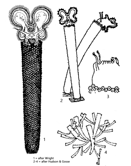
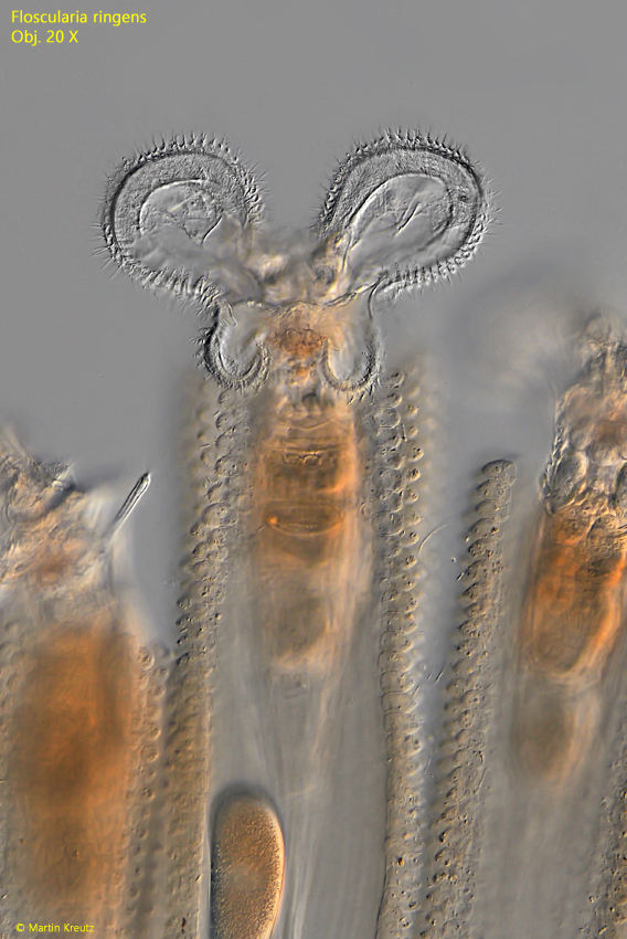
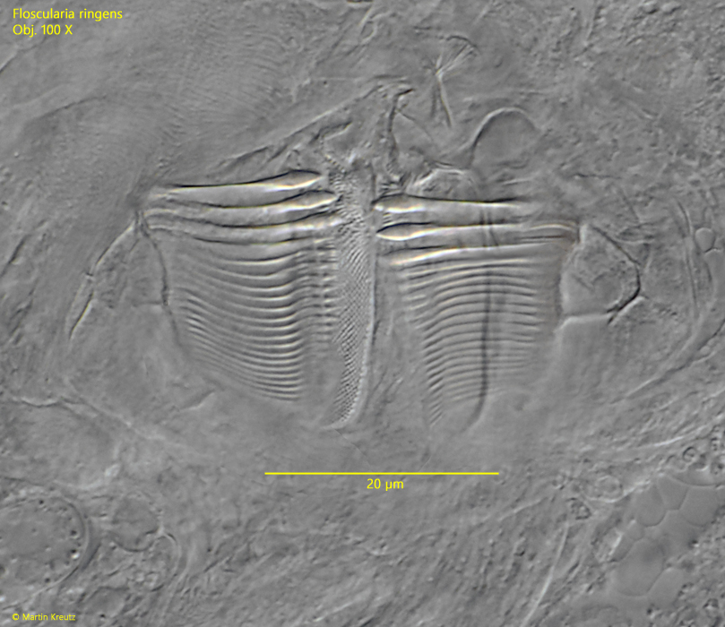
Rotifers of the genus Floscularia can be recognized by their corona, which consists of 4 lobes (s. fig. 4). The genus Limnias has 2 lobes and the genus Ptygura has a circular corona. The tube of Floscularia ringens is built of very regularly arranged pills sitting on a gelatinous membrane. The pills are not formed from fecal balls, but from detritus that has been collected from the environment. On the ventral side, below the corona, Floscularia ringens has a special cavity called modulus, in which the pills are formed.
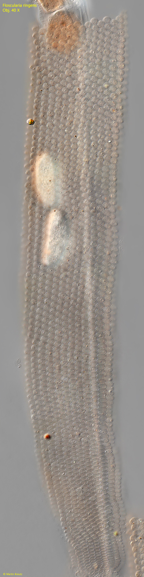
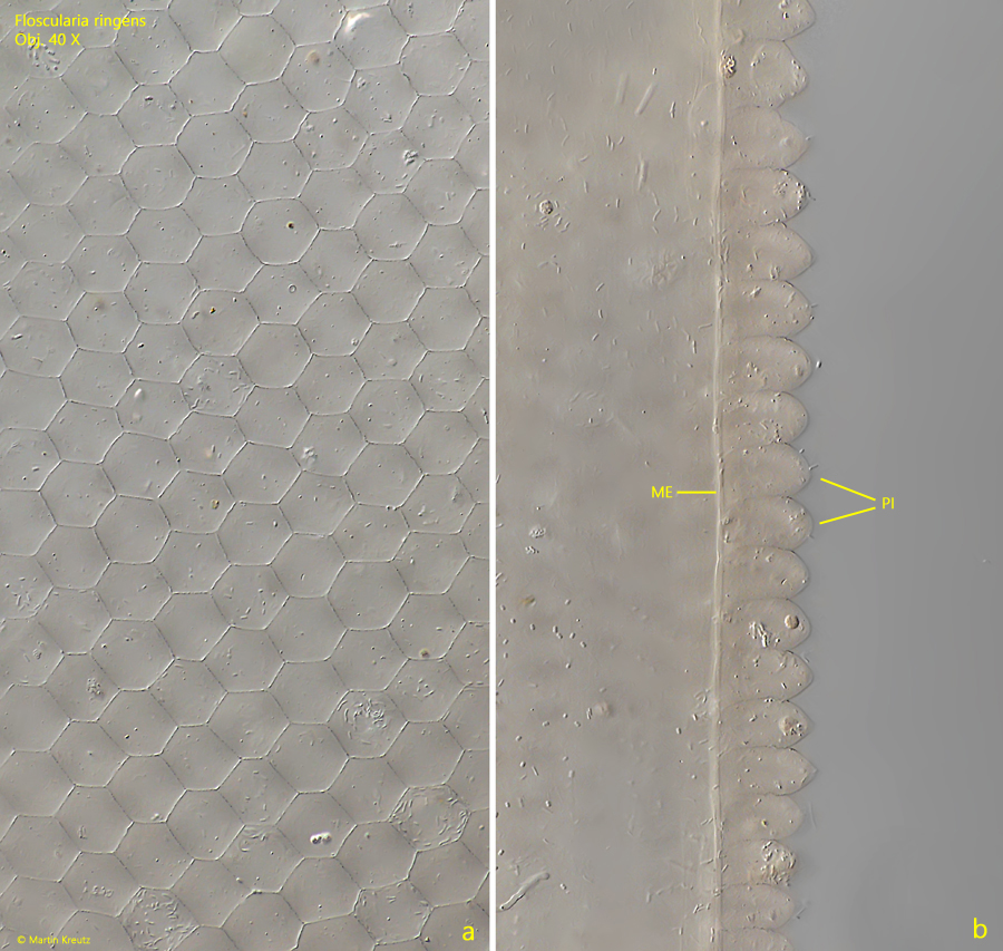
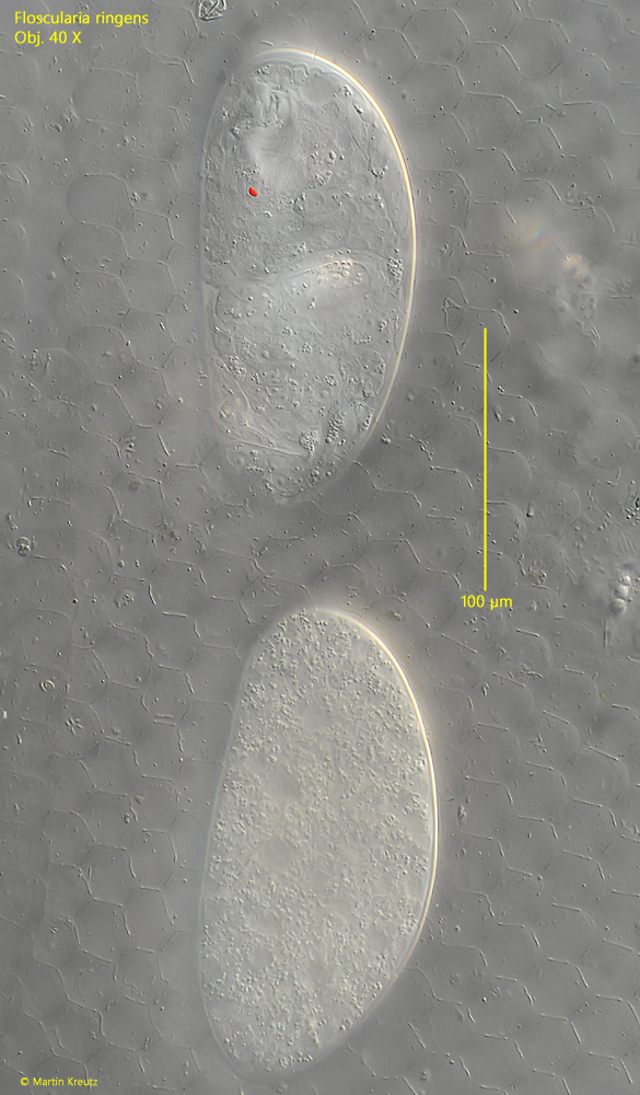
In frontal view, the surface of the tube reveals a very regular hexagonal pattern caused by the densely arranged pills (s. figs. 8 and 9 a). In cross-section, the pills look similar to bullets, with a blunt pointed end (s. fig. 9 b). The color and transparency of the tube is determined by the detritus collected from the environment. The eggs with a smooth surface and a length of about 180 µm are deposited in the tube (s. fig. 10).
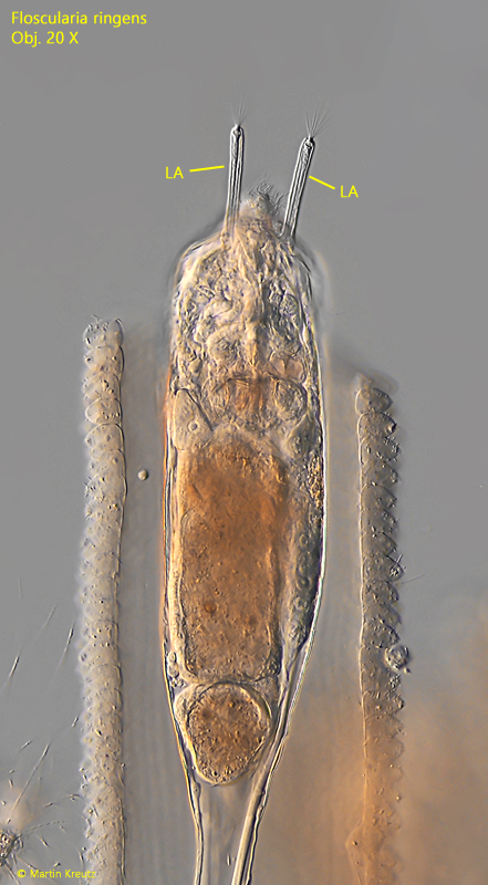
On the ventral side, below the corona, the two lateral antennae are located, which are very long in Floscularia ringens. They have sensory bristles at their distal end, which are connected to a nerve cell. In extended specimens, the corona and lateral antennae protrude from the tube.
Fig. 1:Floscularia ringens. A branched colony of specimens. Obj. 4 X.
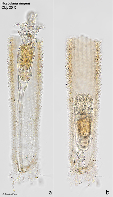
Fig. 2 a-b:Floscularia ringens. L = 690 µm, an elongated (a) and retracted (b) solitary specimen. Obj. 20 X.
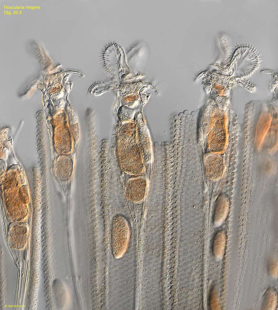
Fig. 3:Floscularia ringens. Some elongated specimens in the branched colony as shown in fig. 1.Obj. 20 X.
Fig. 4:Floscularia ringens. The four lobes of the corona of a fully elongated specimen in ventral view. Obj. 20 X.
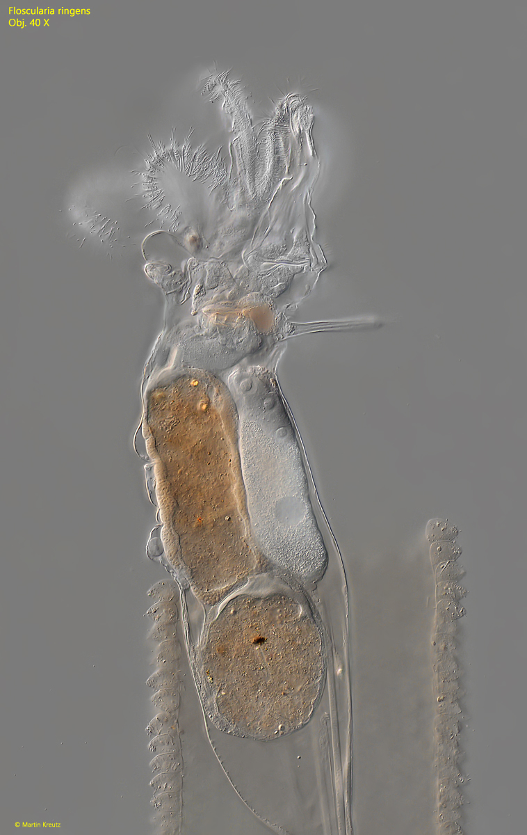
Fig. 5:Floscularia ringens. A partly elongated specimen in lateral view from left. Obj. 40 X.
Fig. 6:Floscularia ringens. The two lateral antennae (LA) of a partly retracted specimen. Obj. 20 X.
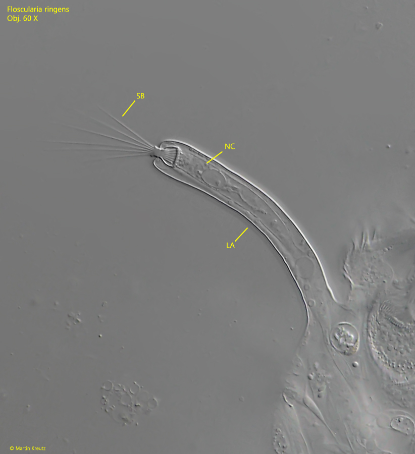
Fig. 7:Floscularia ringens. One of the lateral antennae (LA) in detail. At the distal end a tuft of sensory bristles (SB) are visible. The bristeles are connected with a nerve cell for transmission of any stimuli to the cerebral ganglion. Obj. 60 X.
Fig. 8:Floscularia ringens. Total view of the tube constructed of hundrets of pills. The tube is 1145 µm long.Obj. 40 X.
Fig. 9 a-b:Floscularia ringens. Details of the tube in frontal (a) and in lateral view (b). The pills are arranged accurately in a hexagonal pattern. In lateral view, the membrane (ME), on which the bullet-shaped pills (PI) are glued, is visible. Obj. 40 X.
Fig. 10:Floscularia ringens. Two eggs deposited in the tube. The eggs with a length of 170–180 µm have a smooth surface. Obj. 40 X.
Fig. 11:Floscularia ringens. The trophi in a strongly squashed specimen. Obj. 100 X.