I found large quantities of Geminella mutabilis in the Lauchsee Moor in Austria in June 2024. This green alga forms long, unbranched filaments that float freely.
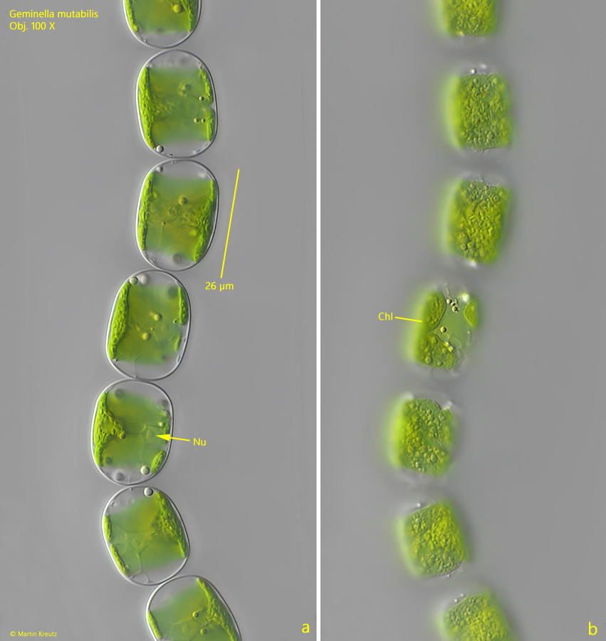
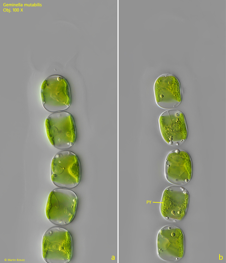
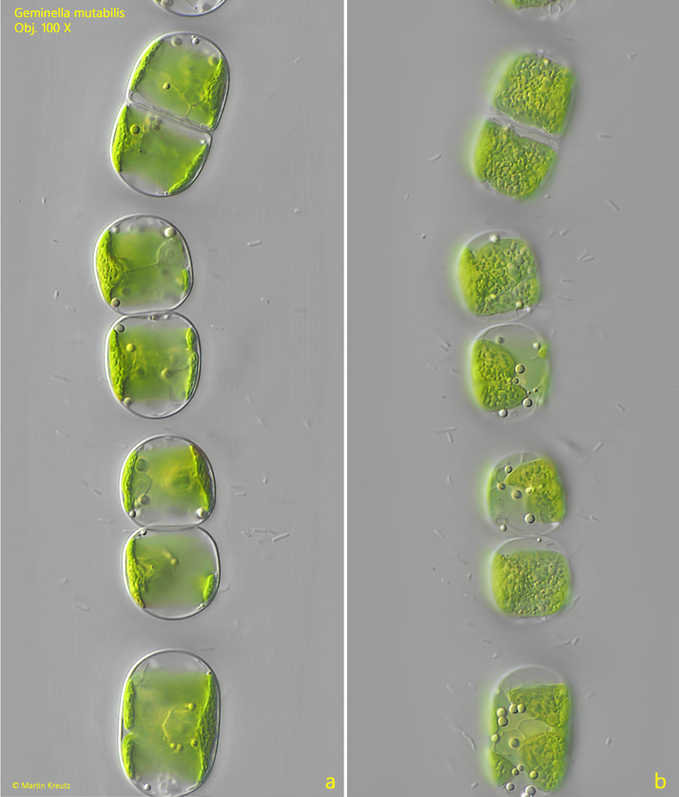
Geminella mutabilis is easily recognizable by its oval cells, which only touch each other slightly at the ends within the filament. The single chloroplast lining the cell wall in the center of the cell is easily recognizable (s. fig. 2 a-b). The cell nucleus is located in the center of the cells (s. fig. 2 a). Young cells are partially vacuolated. The cells become slightly smaller at the cell ends (s. fig. 3 a-b). Division within the filament is also frequently visible there (s. fig. 4 a-b).
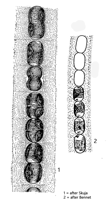
The similar species Geminella minor has much smaller and narrower cells that are rectangular in shape, and in the species Geminella interrupta, there is always a pair of cells together, with a gap to the next pair.
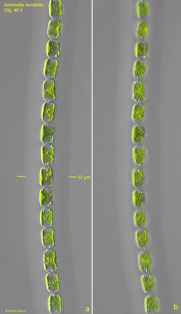
Fig. 1 a-b:Geminella mutabilis. L = 25–28 µm (of cells). Two focal planes of a filament. The filament is covered with a distinct, finely layered gelatinous sheath (d = 53 µm). Obj. 40 X.
Fig. 2 a-b:Geminella mutabilis. L = 23–26 µm (of cells). The oval cells in a filament in detail. The single chloroplast (Chl) lines the cell in a coat-like manner. Nu = nucleus. Obj. 100 X.
Fig. 3 a-b:Geminella mutabilis. L = 20–23 µm (of cells). Two focal planes of the terminal end of a filament. PY = pyrenoid. Obj. 100 X.
Fig. 4 a-b:Geminella mutabilis. A filament with several cells in division. Obj. 100 X.