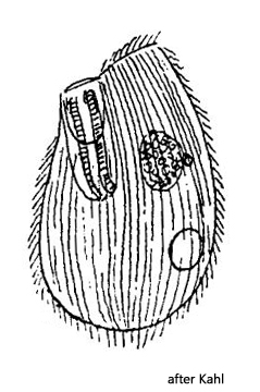
oral apparatus Glaucoma type, undulating membrane right
dense longitudinal rows of cilia
cilia short
contractile vacuole in posterior third
sphaerical macronucleus central
life style between Chaetophora algae
Glaucoma chaetophorae
Glaucoma chaetophorae was first described as Balantiophorus chaetophorae by Penard (1922). Later Kahl (1931) combined the species with Glaucoma.
I found only one specimen of Glaucoma chaetophorae in July 2025 in samples from the Schwemm Moor. Due to its small size, this species is easy to overlook.
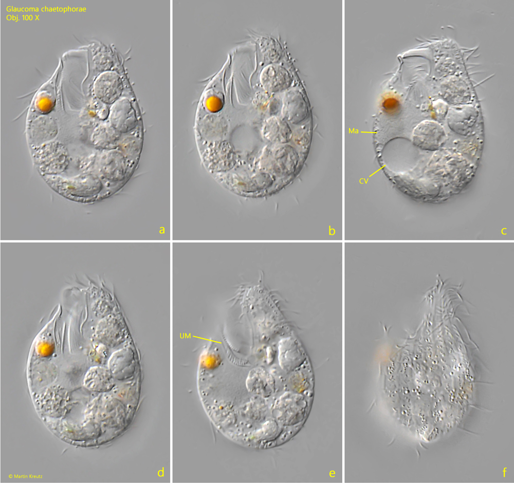
At first glance, the ciliate does not immediately resemble Glaucoma because the mouth opening is apical and laterally shifted. In addition, the oral apparatus is very large, occupying about one-third of the body length. However, in the freely swimming specimen, one can recognize the fan-like movement of the membranelle in the oral cavity, which is typical for Glaucoma.
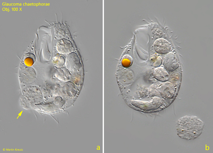
My specimen contained a large number of food vacuoles, which made it difficult to see the central macronucleus (s. fig. 1 c). The contractile vacuole is located in the posterior third, as described by Kahl (s. fig. 1 c). I counted approximately 22–26 longitudinal rows of cilia. The defecation pore is located in the posterior third on the ventral side (s. fig. 2 a-b).
Fig. 1 a-f:Glaucoma chaetophorae. L = 30 µm. Different focal planes of a freely swimming specimen from ventral (a-e) and from dorsal (f). CV = contractile vacuole, Ma = macronucleus, UM = undulating membrane. Obj. 100 X.
Fig. 2 a-b:Glaucoma chaetophorae. L = 30 µm. The same specimen as shown in fig. 1 a-f during the process of defecation (arrow). Obj. 100 X.