oral apparatus subapical, oblique to longitudinal axis
pre-oral suture present (hard to see)
mouth opening with 3 adoral membranelles
undulating membrane right
30–40 longitudinal rows of cilia
macronucleus spherical, mid-body
one adjacent, sphaerical micronucleus
contractile in posterior third
one excretion pore dorsal
some elongated caudal cilia
Glaucoma scintillans
Glaucoma scintillans is a very common ciliate that is particularly common in nutrient-rich habitats with high bacterial densities. It is also regularly found in hay infusions.
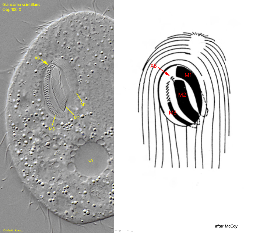
Although Glauoma scintillans is relatively small, measuring approximately 50-60 µm in length, it is noticeable in samples due to the fanning activity of the membranelles in the mouth opening. These move at a frequency of 7–10 Hz, which is easily visible to the human eye. The mouth opening itself is vertical and parallel to the longitudinal axis of the body, but the adoral membrane and the undulating membrane are slightly slanted, which is typical for the genus Glaucoma. Upon closer inspection of the mouth opening, the three adoral membranelles are clearly visible (s. fig. 4). At the anterior end of membranelle 2, there is also a small, slightly offset ciliated field called the X-body (s. fig. 4).
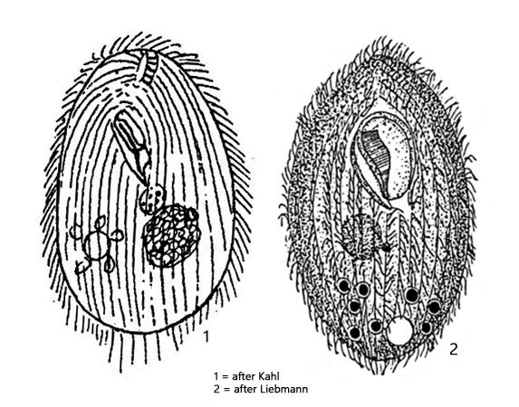
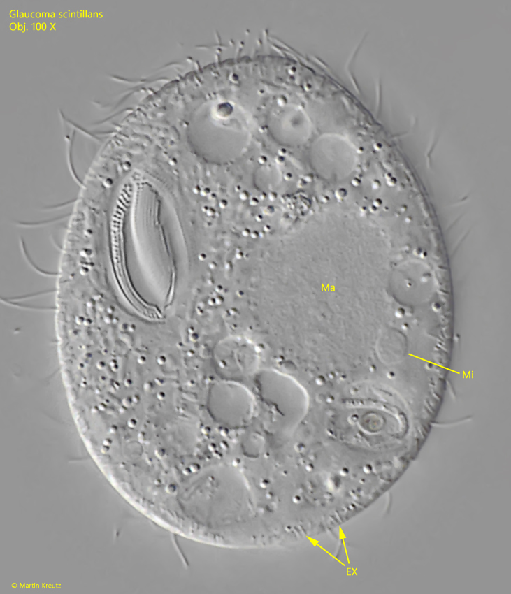
The food of Glaucoma scintillans appears to consist exclusively of bacteria found in all food vacuoles. The globular macronucleus with attached micronucleus is located in the center of the body (s. fig. 6). The contractile vacuole is located at the border to the posterior third (s. figs. 3 b and 4) and was drawn too far shifted posterior by Liebmann (s. drawing 2, above).
To my knowledge, the very small extrusomes, which are located benenath to the pellicle, have not yet been described in detail. According to my observations, they are rod-shaped and have a length of 1.5 µm (s. fig. 6).
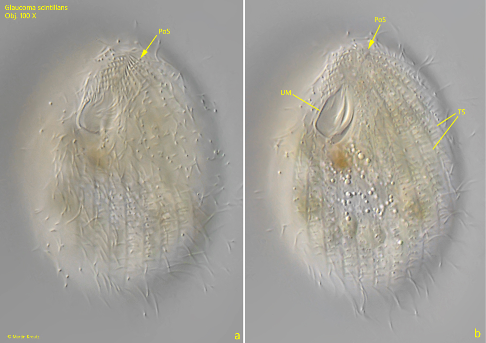
In some specimens of Glaucoma scintillans, I was able to detect transverse striations on the pellicle, which are also sometimes found in other species of the genus Glaucoma (s. fig. 5 a-b). However, these transverse striations did not appear to be equally pronounced in all specimens. In some specimens, they seemed to be absent.
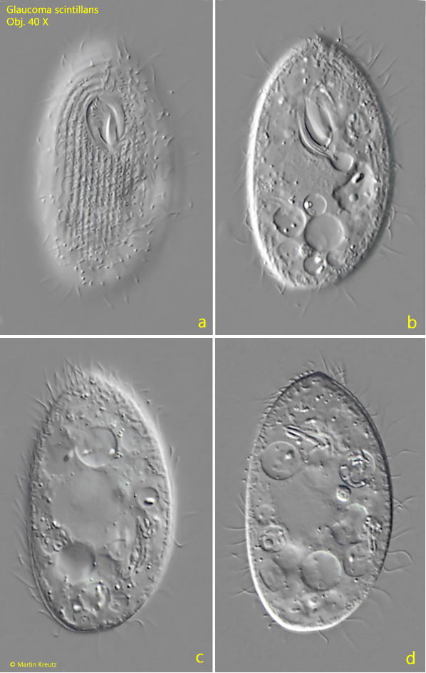
Fig. 1 a-d:Glaucoma scintillans. L = 57 µm. Different focal planes of a freely swimming specimen from ventral. Obj. 40 X.
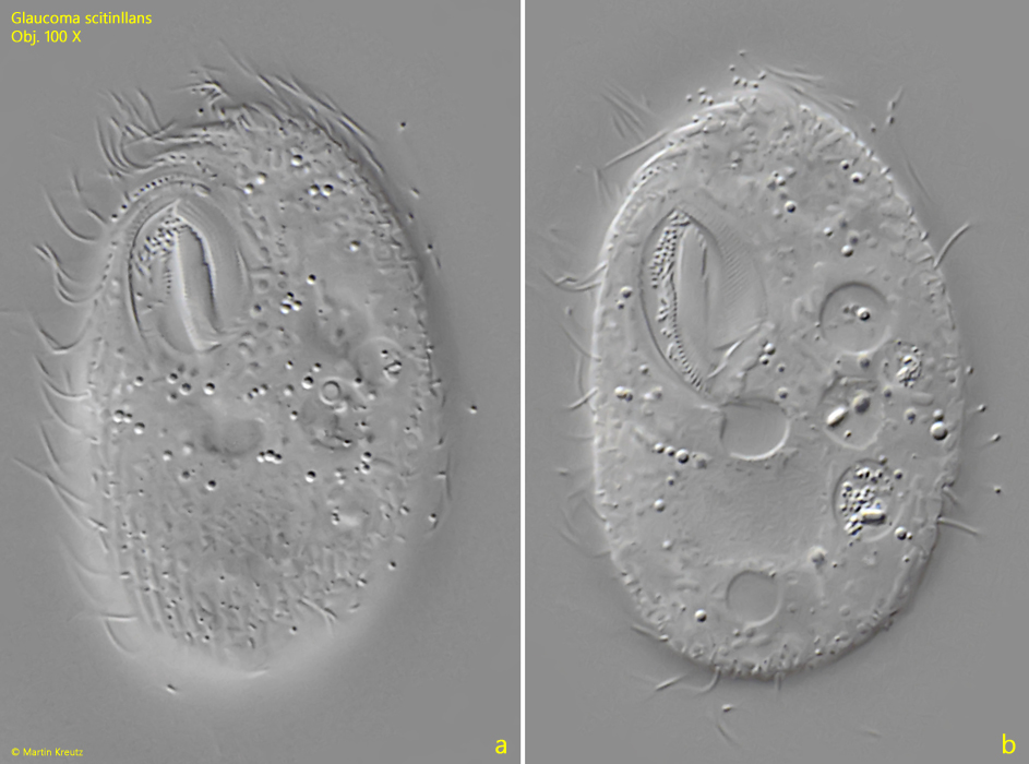
Fig. 2 a-b:Glaucoma scintillans. L = 48 µm. A second freely specimen from ventral in detail. Obj. 100 X.
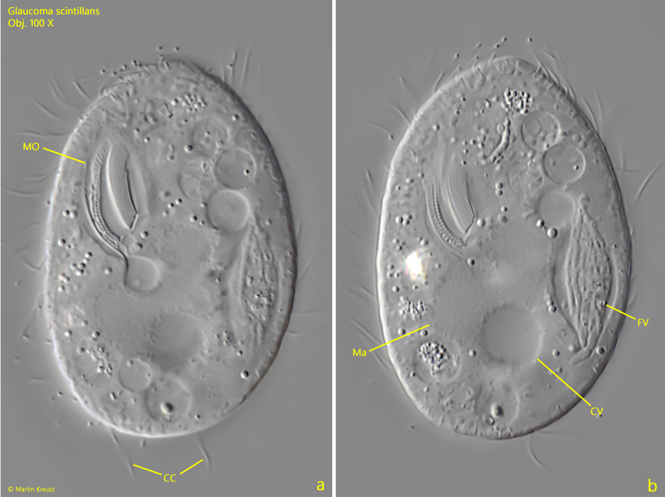
Fig. 3 a-b:Glaucoma scintillans. L = 47 µm. A slightly squashed specimen from ventral. The food vacuole (FV) on the left side is filled with a bundle of bacteria. CC = elongated caudal cilia, CV = contractile vacuole, Ma = macronucleus, MO = mouth opening. Obj. 100 X.
Fig. 4:Glaucoma scintillans. The oral apparatus in detail compared to a schematic drawing. The three adoral membranelles alles visible (M1–M3) and the so called X-body (XB), a small field of cilia at the anterior end of the membranelle M2. CV = contractile vacuole. Obj. 100 X.
Fig. 5 a-b:Glaucoma scintillans. L = 53 µm. The pre-oral suture (PoS) in a freely swimming specimen. Note the delicated transverse striation (TS) of the pellicle. On the right side of the mouth opening the undulating membrane (UM) is visible. Obj. 100 X.
Fig. 6:Glaucoma scintillans. The macronucleus (Ma) and the adjacent micronucleus (Mi) in a squashed specimen. Beneath the pellicle the rod-shaped extrusomes (EX) are visible with a length of 1.5 µm. Obj. 100 X.
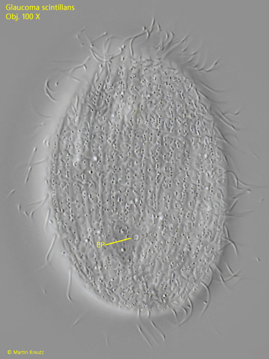
Fig. 7:Glaucoma scintillans. The single excretion pore (EP) of the contractile vacuole on the dorsal side. Obj. 100 X.