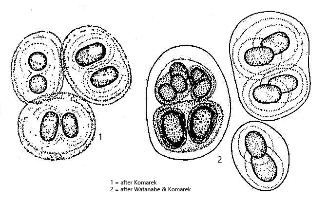
cells covered by colorless, yellow or brownish sheath
sheath often contains finely granular inclusions
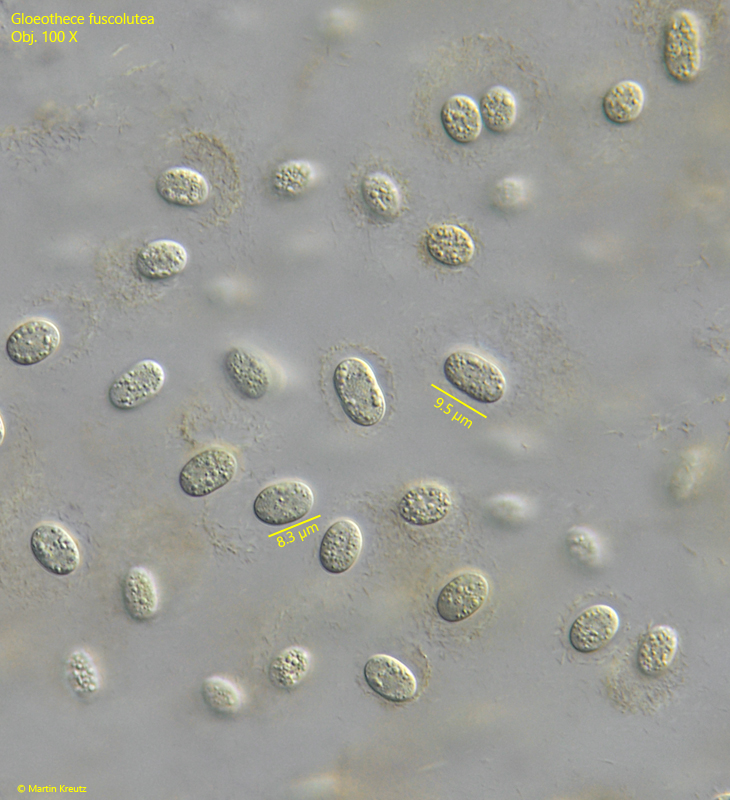
Gloeothece fuscolutea
The colonies of the cyanobacterium Gloeothece fuscolutea can be recognized by their yellowish to yellow-brown color. This is caused by a precipitate that permeates the mucilaginous envelopes of the individual cells and is probably produced by the cells themselves. It does not appear to be iron deposits, which are often found in the mucilaginous envelopes of green algae. This secretion may serve as UV light protection, as I found the colonies in shallow and almost dried-out puddles in the Sima Moor.
The also yellow-brown colored similar species Gloeothece rupestris differs mainly in the size of the cells and habitat. The cells of Gloeothece rupestris are only half as large, and the colonies occur terrestrially on wet rocks.
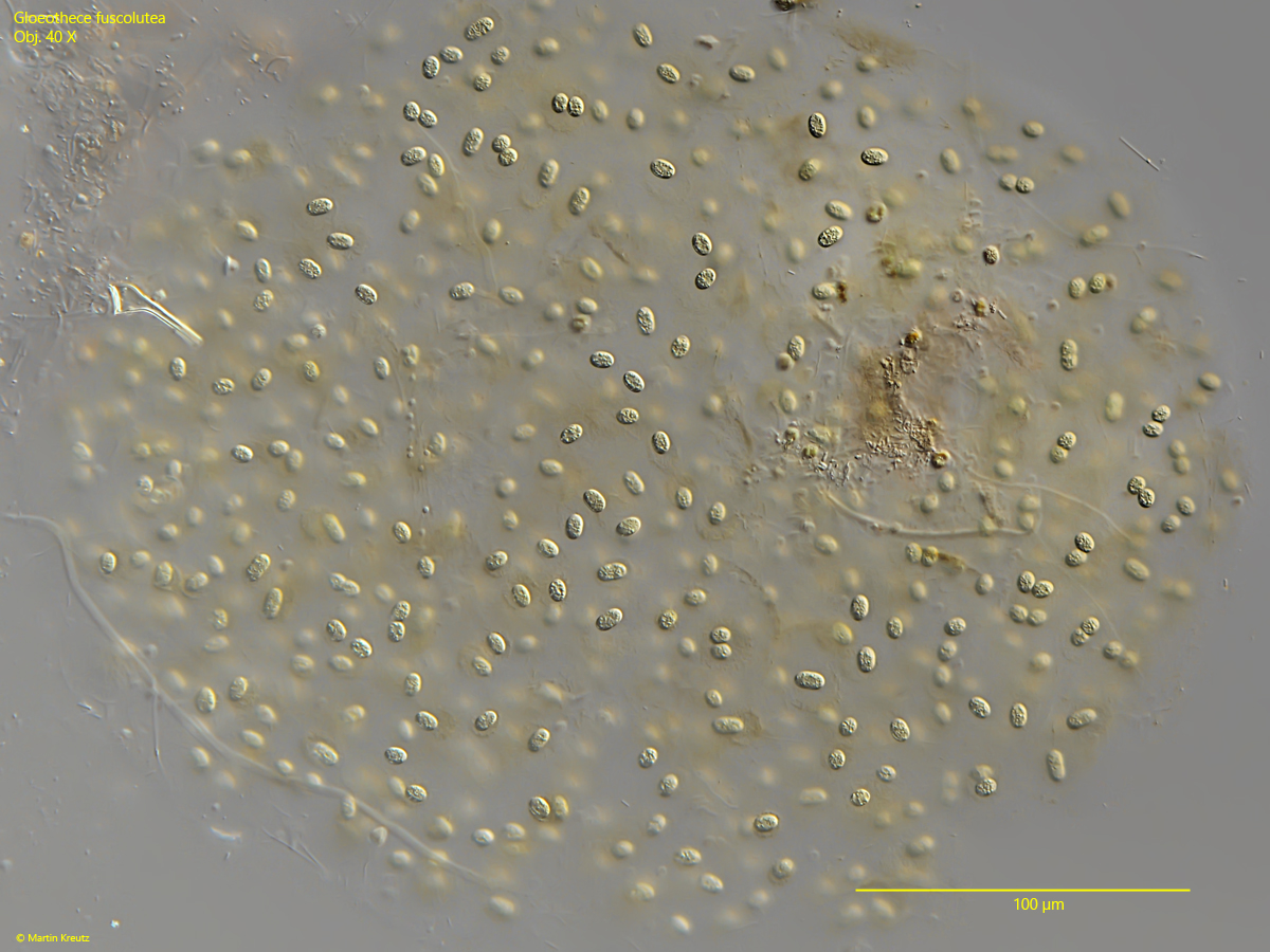
Fig. 1:Gloeothece fuscolutea. L = 380 µm (of colony). A yellowish colored colony found in June 2025 in the Sima Moor. Obj. 40 X.
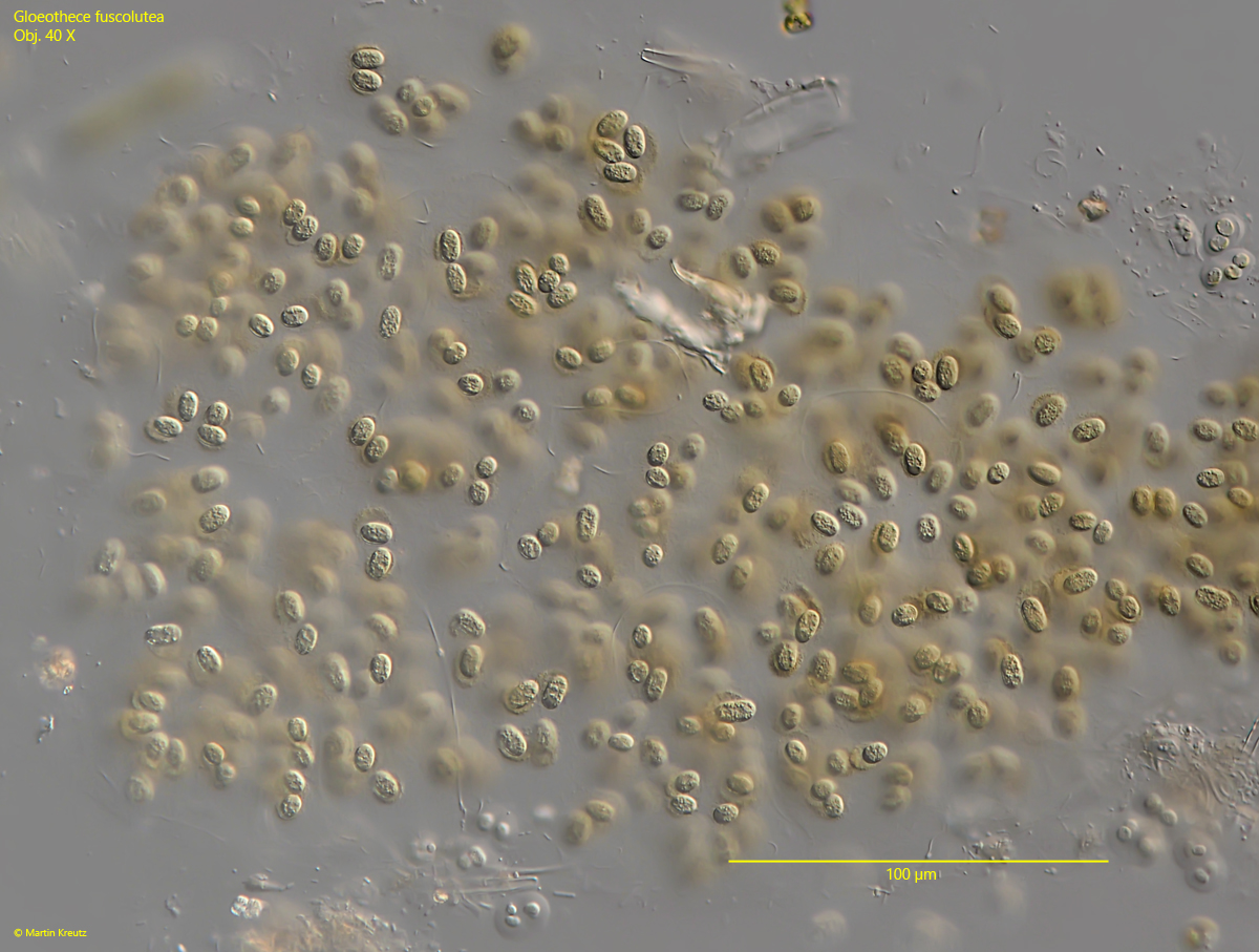
Fig. 2:Gloeothece fuscolutea. L = 320 µm (of colony). A second, more brownish colored colony. Obj. 40 X.
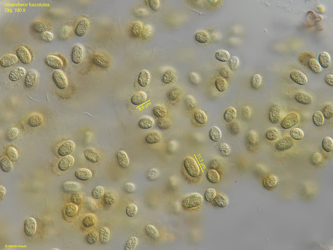
Fig. 3:Gloeothece fuscolutea. L = 8–11.5 µm (of cells). Some cells in the colony as shown in fig. 2. The mucilaginous sheath of the cells contain a yellow-brownish precipitation. Obj. 100 X.
Fig. 4:Gloeothece fuscolutea. L = 8–10 µm (of cells). Some cells in the colony as shown in fig. 1. The mucilaginous sheath of the cells contain only a small amount of a yellow-brownish precipitation. Obj. 100 X.