caudal ventral pair of spines exceeding body and cross each other
dorsolaterally five pairs of curved spines
ventral side with 2 rows of cilia in 13–14 groups
Haltidytes crassus
I regularly find Haltidytes crassus in the mud of Simmelried and the Purren pond. The specimens are easy to recognize by the long caudal spines, which are significantly longer than the body and cross at the back. The body itself appears compact and bottle-shaped. The head is clearly set off from the body and covered with long cilia, which also serve for locomotion.
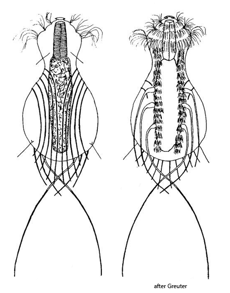
On the ventral side, three pairs of spines arise. The last, rearmost pair consists of the long spines that exceed the body length. According to my observations, the two anterior pairs consist of double spines (s. fig. 4 a), which differs from the drawings by Greuter (s. above) and the descriptions by Schwank (1990). However, this arrangement of the ventral spines was also made by Müller (2023, s. link below).
Dorsolaterally, 5 pairs of simple spines arise, which exceed the body by 20–30 µm. Whether they are involved in an escape movement is very difficult to determine. In any case, the long ventral spines serve a very fast, jumping escape movement.
In the specimens of my population, the body was always over 100 µm long. The largest specimens measured 125 µm. That is about 15% longer than stated by Schwank.
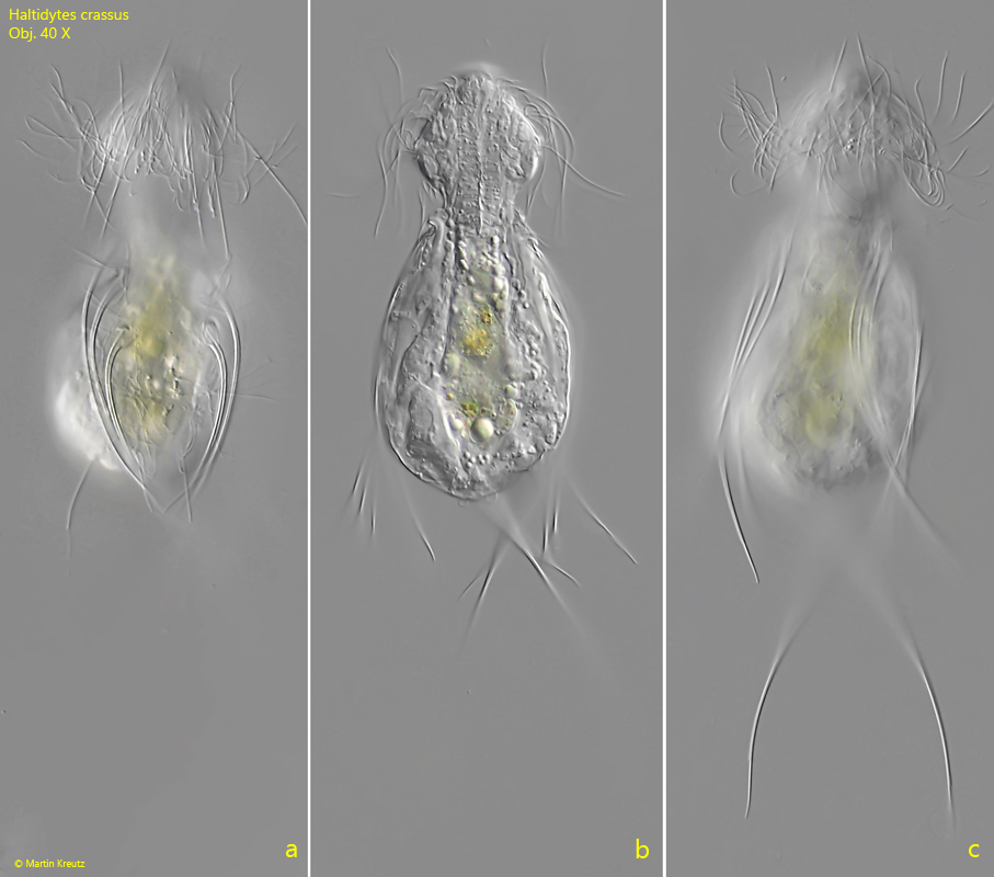
Fig. 1 a-c:Haltidytes crassus. L = 107 µm (of body). A freely swimming specimen from ventral. The 3 pairs of ventral spines are visible (a). Obj. 40 X.
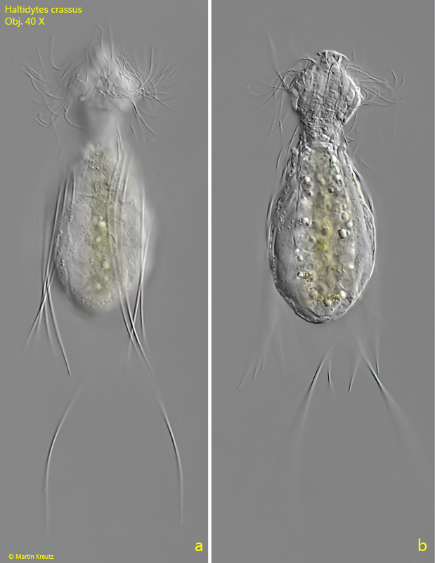
Fig. 2 a-b:Haltidytes crassus. L = 113 µm (of body). A slightly squashed specimen from dorsal. The five pairs of spines are visible (1-5) which arise dorsolaterally. Obj. 40 X.
Fig. 3 a-b:Haltidytes crassus. L = 124 µm (of body). A second specimen from dorsal. Obj. 40 X.
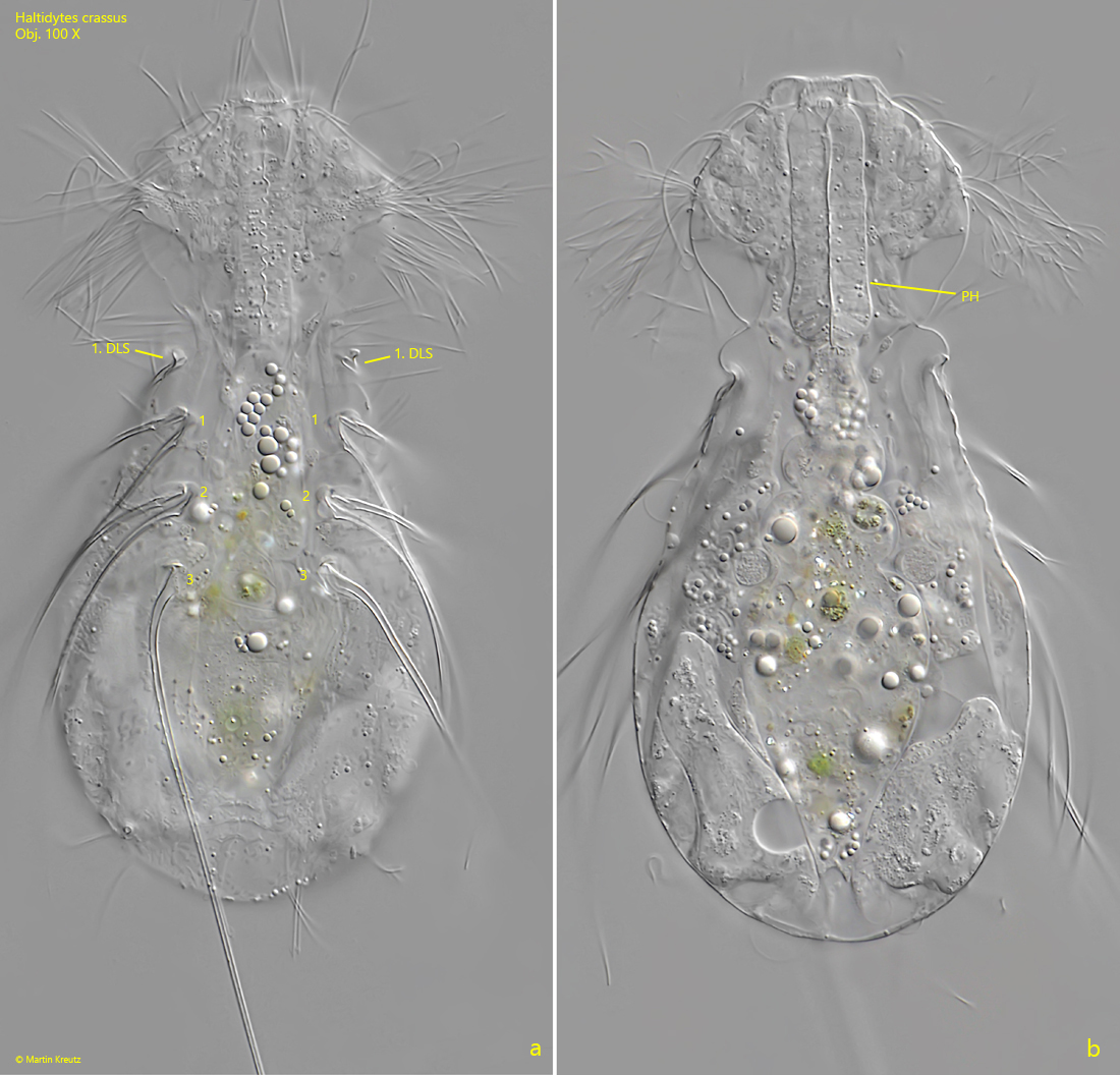
Fig. 4 a-b:Haltidytes crassus. Two focal planes of a sqaushed specimen from ventral. The three pairs of ventral spines are vsible. The pairs 1 and 2 are consisting of double spines. In the neck region the first pair of dorsolateral spines (1. DLS) is visible. PH = pharynx. Obj. 100 X.
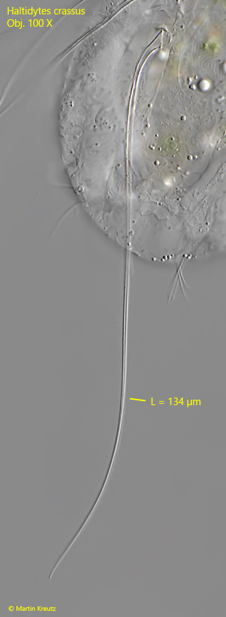
Fig. 5:Haltidytes crassus. The caudal spines on the ventral side exceeding the body and cross each other. This is the right caudal spine with a length of 134 µm. Obj. 100 X.