I found Hastatella aeculacantha in August 2026 in the heavily eutrophic pond of the waste disposal company Constance. The specimens were found only sporadically in the plankton samples. However, in the sample containers, the specimens accumulated at the water surface. There, the specimens move in slow circles.
Hastatella aesculacantha has so far been found only rarely. What all sites have in common is that they were heavily eutrophic. Jarocki (1927) reported the ciliates Phascolodon vorticella and Actinobolina radians as accompanying fauna. I was unable to detect Phascolodon vorticella in my samples, but I did find Actinobolina radians. In addition, specimens of Linostomella vorticella were present.
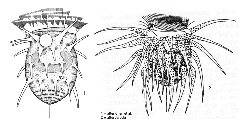
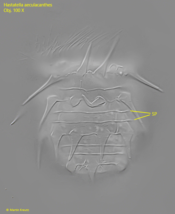
The most striking feature of Hastatella aesculacantha is the epiplasmic spines covering its body. Their length is evidently subject to considerable variation. Jarocki (1927) illustrated them as very long, whereas Chen et al. (2022), who studied a Chinese population, illustrated the spines as considerably shorter (s. drawings above). The specimens of my population more closely resembled the Chinese population.
In Hastatella aesculacantha, the spines are arranged in 4 transverse rows. This can best be recognized in squashed specimens (s. fig. 5 a). When the ciliate is disturbed, the peristome immediately retracts. As a result, the body becomes rounded and the spines spread apart. When the peristome is extended again, the spines lie flat against the body. The pellicle has very broad striation, which is clearly visible in slightly squashed specimens (s. fig. 4). There are said to be about 15 striations, which become increasingly dense toward the posterior end. At the posterior end, two rigid bristles are described, which I was unable to detect, however.
The contractile vacuole lies directly at the anterior margin, on the dorsal side of the infundibulum (s. fig. 2 a). The macronucleus is C-shaped and lies in the anterior third (s. fig. 5 b). I was unable to detect a micronucleus. However, it was also not described by earlier authors.
Hastatella aesculacantha can be distinguished from the similar species Hastatella radians by its 4 rows of spines. Hastatella radians possesses only 2 rows of spines.