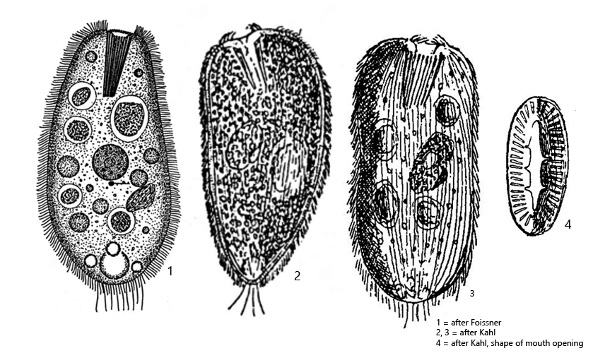
body long oval or cylindrical, anteriorly often slightly obliquely truncated
cytoplasm transparent, symbiotic algae absent
length 80–300 µm (usually 150–250 µm)
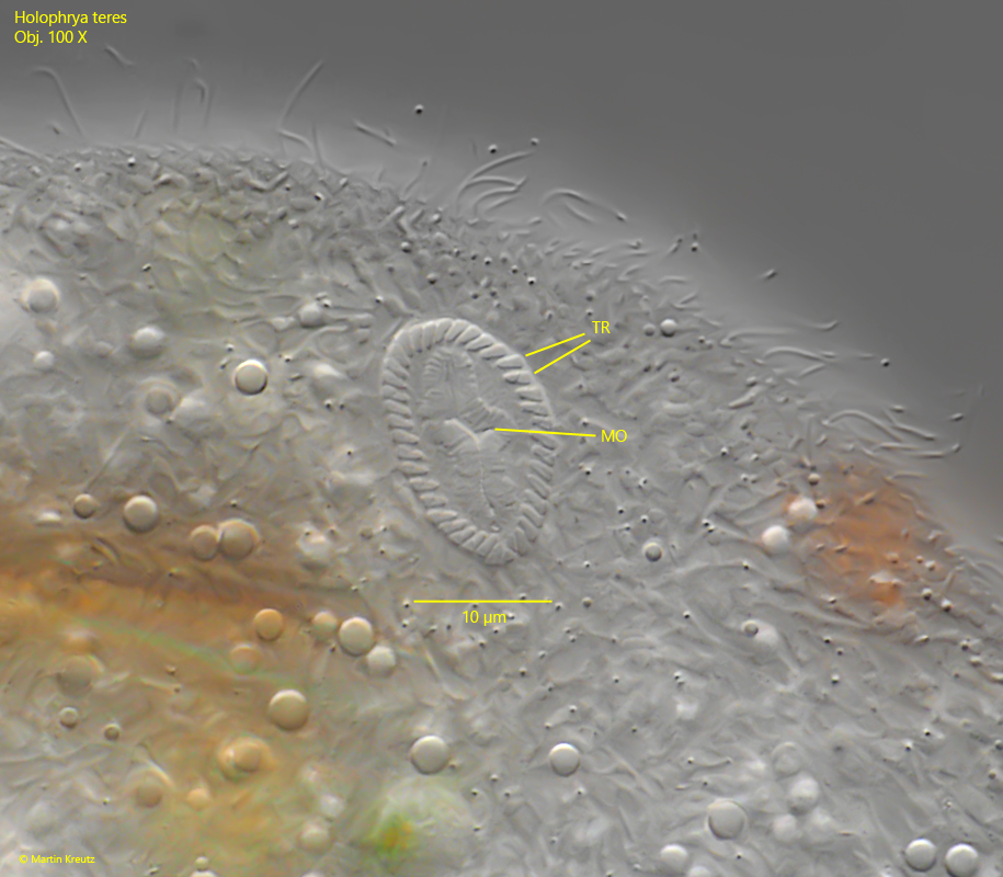
mouth opening apical, oral basket with about 40–50 trichites
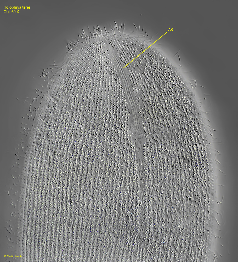
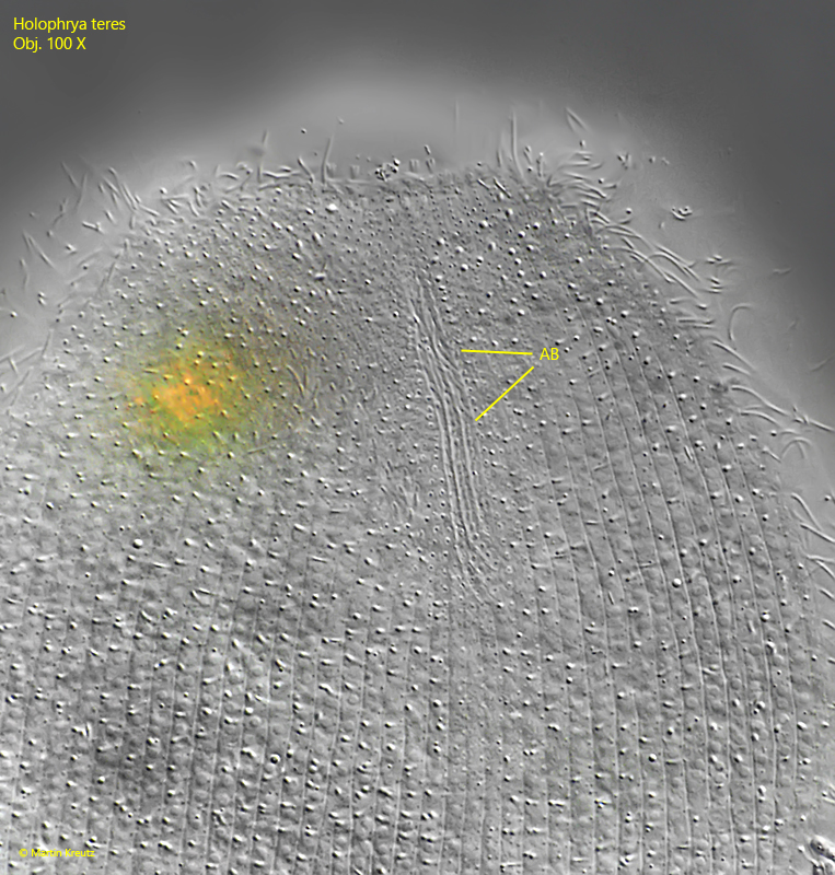
adoral brush (3 rows), in dexiotropic position
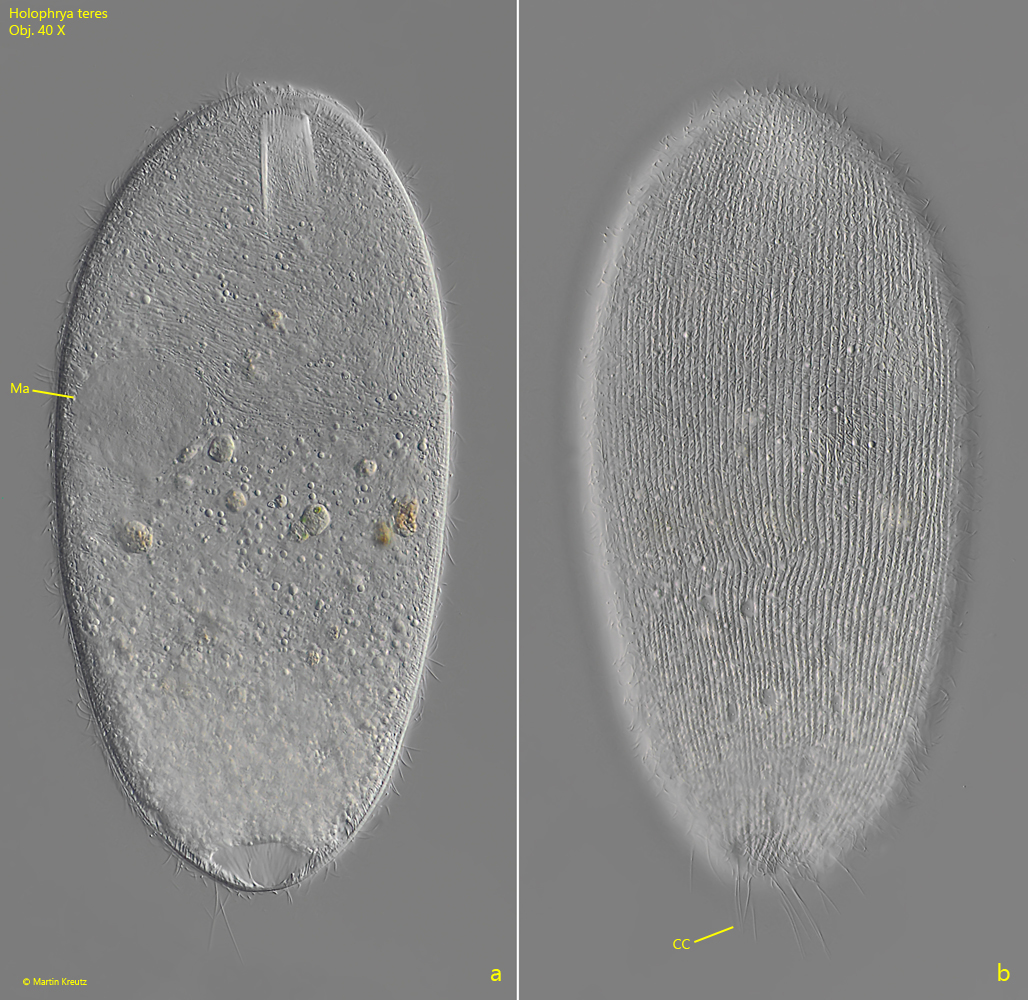
80–110 longitudinal rows of cilia
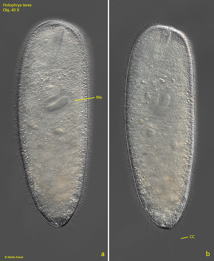
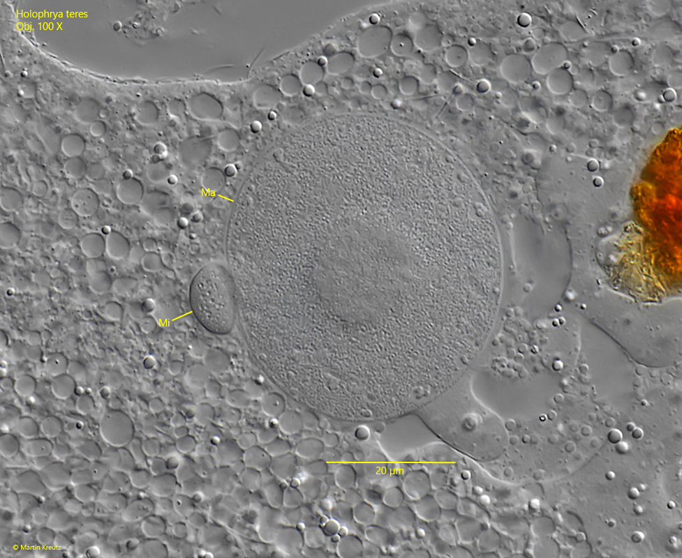
macronucleus ellipsoid, one adjacent large micronucleus
extrusomes short, rod- or spindel-shaped
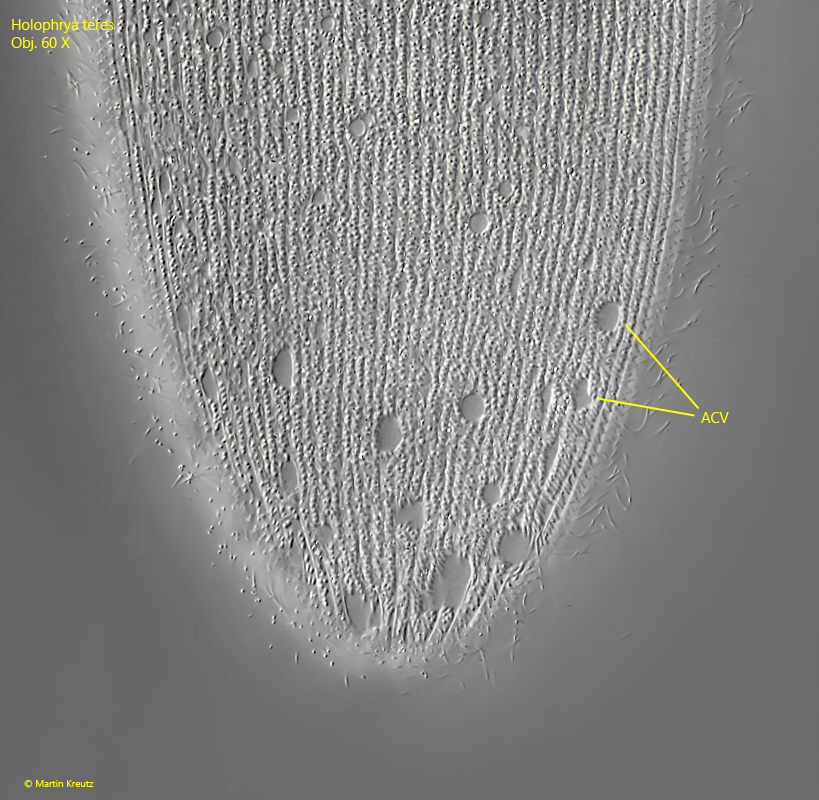
contractile vacuole terminal with numerous auxiliary vacuoles
several caudal cilia
Holophrya teres
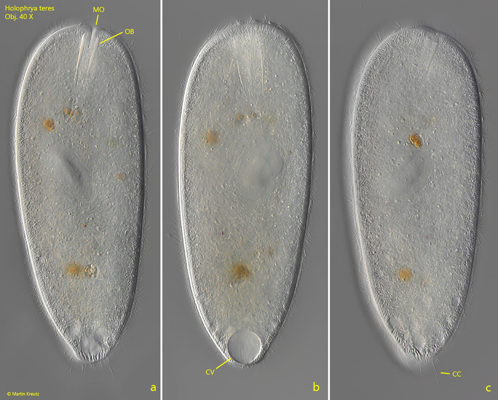
Holophrya teres is a very common ciliate that occurs in almost all of my sampling sites. The species has a complex life cycle (theront-trophont-protomont-tomont-tomit). The images below show the theront phase, with little ingested food. In the trophont and protomont phases, the specimens are completely filled with food vacuoles, rounded and opaque.
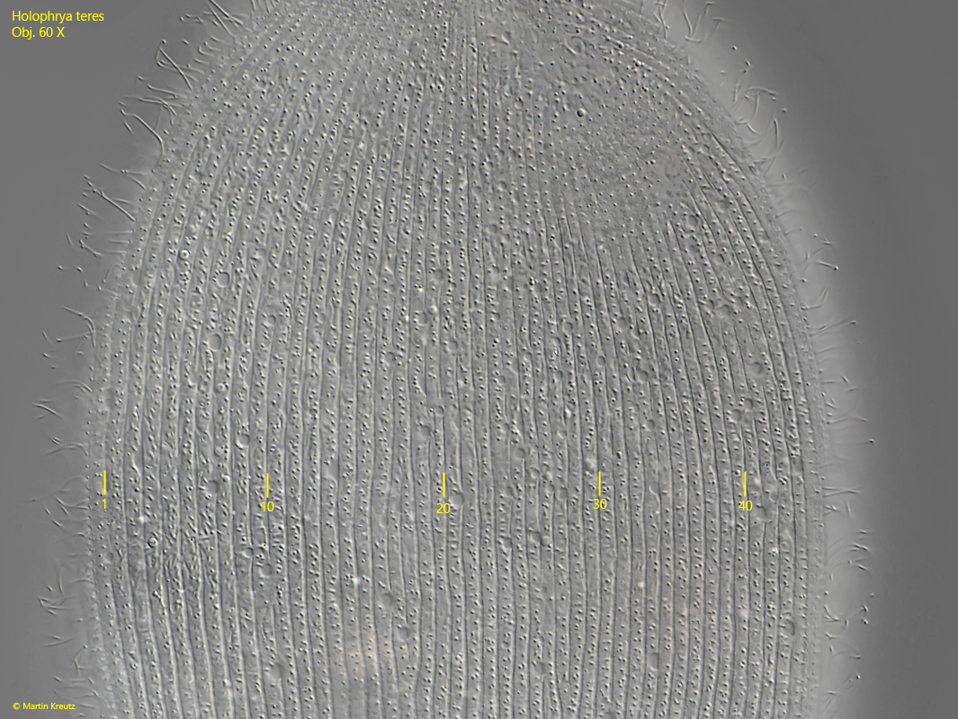
Important characteristics for the identification are the number of longitudinal rows of cilia, the size and the absence of symbiotic algae. The cell surface is covered by 80–110 longitudinal rows of cilia, which are located in furrows. The total number can only be seen in anterior or posterior views. In lateral view, about 40–50 longitudinal rows can be recognized in squashed specimens (s. fig. 4). Another characteristic feature of Holophrya teres is the shape of the mouth opening (s. drawing 4, above). The trichites of the oral basket are not arranged in a circle, but in an oval (s. fig. 8) or sometimes almost rectangular.
The similar species Holophrya discolor is smaller (usually 80–120 µm) and has only 35–64 longitudinal rows, i.e. about half as many as Holophrya teres. If the cytoplasm contains symbiotic algae and the cells are smaller than 160 µm, then it is Holophrya ovum.
In my population, the specimens of Holophrya teres were consistently longer than 250 µm. The oral basket consisted of about 40 trichites in most specimens. All specimens had about 20–30 caudal cilia. I had the impression that these were arranged in a circle around the posterior cell pole (s. figs. 3 a and 3 b).
Fig. 1 a-c:Holophrya teres. L = 270 µm. A freely swimming specimen. CC = caudal cilia, CV = contractile vacuole, MO = mouth opening, OB = oral basket. Obj. 40 X.
Fig. 2 a-c:Holophrya teres. L = 282 µm. A second, freely swimming specimen. CC = caudal cilia, Ma = macronucleus. Obj. 40 X.
Fig. 3 a-b:Holophrya teres. L = 280 µm. Two focal planes of a slightly squashed specimen. CC = caudal cilia, Ma = macronucleus. Obj. 40 X.
Fig. 4:Holophrya teres. The longitudinal rows of cilia in a squashed specimen. On this side of the body 46 rows are visible. Obj. 60 X.
Fig. 5:Holophrya teres. The adoral brush (AB) of a slightly squashed specimen. Obj. 60 X.
Fig. 6:Holophrya teres. The adoral brush (AB) of a second, squashed specimen. Obj. 100 X.
Fig. 7:Holophrya teres. Beneath the pellicle numerous auxiliary contractile vacuoles are located, especially in the posterior fifth of the body. They are connected to the terminal contractile vacuole via collecting canals. Obj. 60 X.
Fig. 8:Holophrya teres. Apical view of the mouth opening (MO) with the 40 trichites of the oral basket (OB). Obj. 100 X.
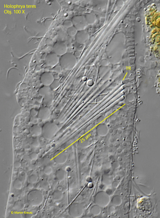
Fig. 9:Holophrya teres. The trichites (TR) of the oral basket about 25 µm. The anterior end is thickended with a tooth. Obj. 100 X.
Fig. 10:Holophrya teres. The macronucleus (Ma) with the adjacent micronucleus (Mi) in a strongly squashed specimen. Obj. 100 X.
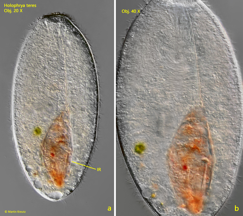
Fig. 11 a-b:Holophrya teres (L = 298 µm). A specimen with an ingested rotifer of the genus Trichocerca. Obj. 20 X (a) and 40 X (b).