body elongated elliptical, dorso-ventrally flattened
length 60–90 µm, width 20–30 µm
2 macronuclei with each one micronucleus
contractile vacuole below equator, left margin
nuclear apparatus
adoral zone of membranelles with apical interruption
undulating membrane curved
3 frontal cirri
1 buccal cirrus
2 frontoterminal cirri
2 rows of ventral cirri
7-12 J-shaped arranged transversal cirri
left and right marginal rows of cirri
dorsally 4 rows of bristle-shaped cilia
caudal cirri absent
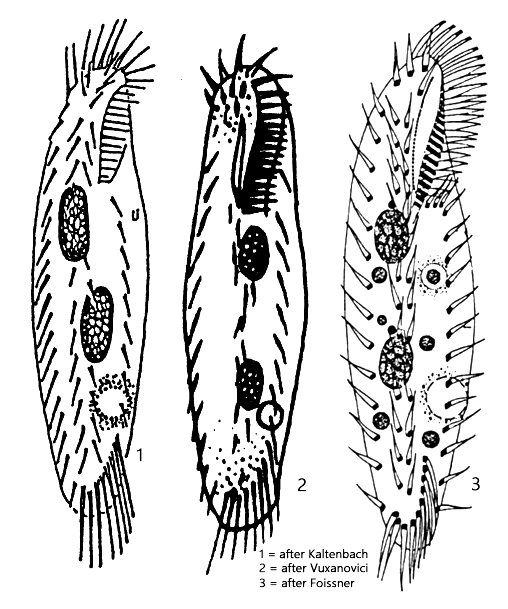
Holosticha pullaster
Holosticha pullaster is one of the most common hypotrich ciliates, found in almost all of my sampling sites. In older samples, specimens can often be found on the surface. The species likes to settle on the floating coverslip and is then easy to observe.
Freely swimming specimens stand out even at low magnifications due to their shape. In slightly contracted specimens, the left side of the body, where the contractile vacuole is also located, is more strongly convexly curved than the right side, which is usually straight. In addition, the contractile vacuole is clearly located below the cell equator. This is also an important distinguishing feature from the similar species Tachysoma pellionellum, whose contractile vacuole is located in the middle on the left side.
The ciliation of Holosticha pullaster essentially consists of a double row of ventral cirri, with a row of marginal cirri on both the right and left sides (s. fig. 2 a-c). The transverse cirri are arranged in the shape of a “J”. The adoral zone has a length of one-quarter to one-third of the body length. On the right side of the mouth opening lies the inconspicuous, curved undulating membrane. There are 7–8 frontal cirri present, which are hard to see.
The nuclear apparatus consists of two oval or elliptical macronuclei, each with an attached micronucleus (s. fig. 3). The macronuclei are always clearly separated from each other. In some specimens, constrictions can be seen on the macronuclei (s. fig. 1 c). These are so-called replication bands. They serve to prepare the macronucleus for an imminent cell division.
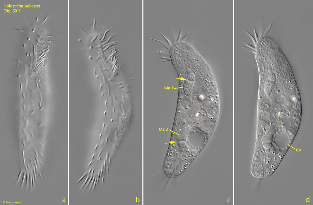
Fig. 1 a-d:Holosticha pullaster. L = 95 µm. Different focal planes of a freely swimming specimen from ventral. The macronuclei (Ma 1, Ma 2) of this specimen have developed so-called replication bands (arrows). CV = contractile vacuole. Obj. 60 X.
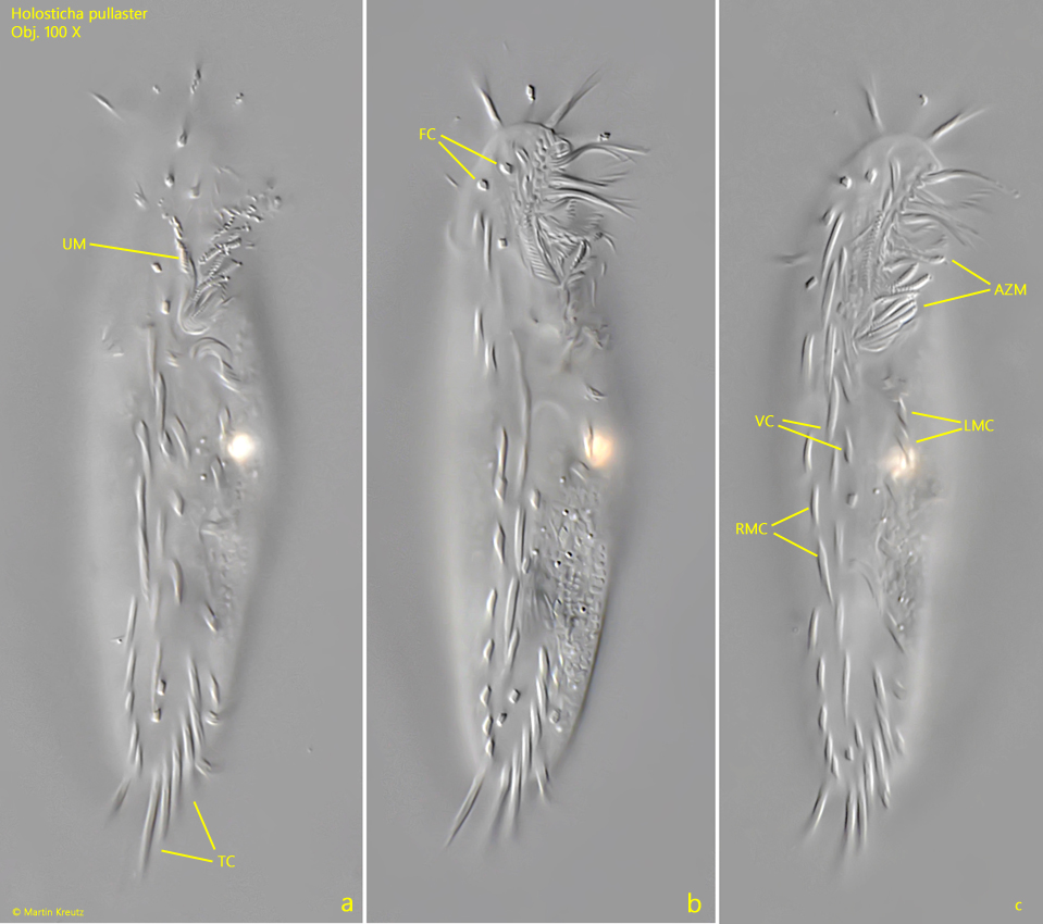
Fig. 2 a-c:Holosticha pullaster. L = 92 µm. The ventral ciliation of a second specimen in detail. There are two middle longitudinal rows of ventral cirri (VC) with each one marginal row of cirri on the left side (LMC) and on the right side (RMC). The undulating membrane (UM) is inconspicuous and curved. The adoral zone of membranelles has about one fouth or one thirds of body length. The transverse cirri (TC) are arranged J-shaped. FC = frontal cirri. Obj. 100 X.
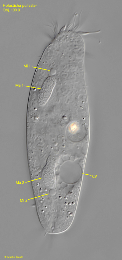
Fig. 3:Holosticha pullaster. L = 91 µm. Focal planes from ventral on the two macronuclei (Ma 1, Ma 2) with each one adjacent micronucleus (Mi 1, Mi 2). Note the contractile vacuole (CV) on the left side clearly below the cell equator. Obj. 100 X.
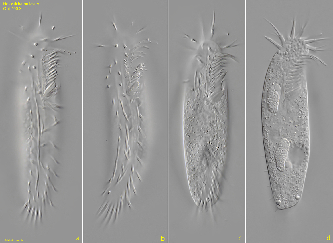
Fig. 4 a-d:Holosticha pullaster. L = 74 µm. Different focal planes of a third specimen from ventral. Obj. 100 X.