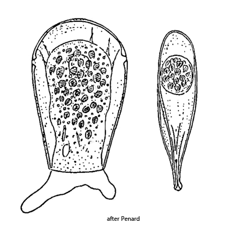
test laterally flattended, wedge-shaped in lateral view
length 70–140 µm
aperture a narrow, elliptical slit, surrounded by thickened lip
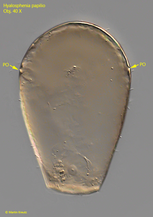
two lateral pores in posterior third of test (hard to see)
spherical nucleus in posterior third of cell
several contractile vacuoles in posterior half of cell
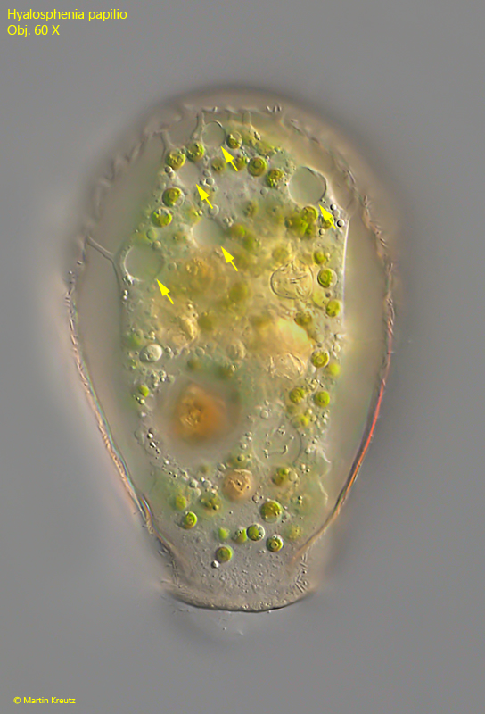
cytoplasm greenish due to symbiotic algae
protoplast attached to test by filaments of cytoplasm
several active pseudopods
Hyalosphenia papilio
So far I have only found Hyalosphenia papilio in the Sima Moor (Austria), where this testate amoeba is very common.
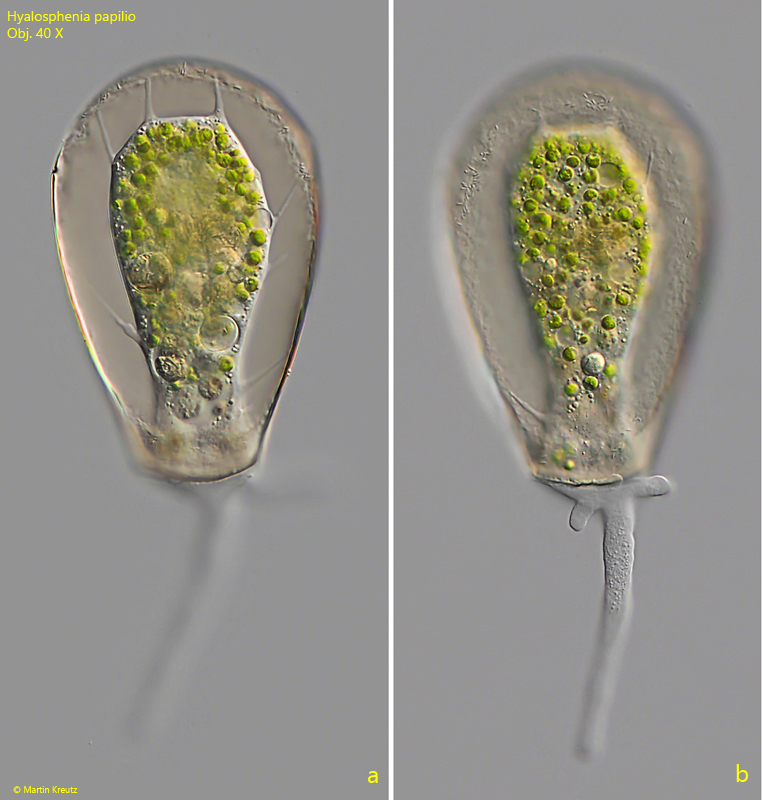
Under the coverslip, Hyalosphenia papilio is usually visible in frontal view. The test then appears pouch-shaped. It is transparent, mostly brownish and without any visible structure. The protoplast is attached to the inside of the test with fine filaments (s. fig. 2). The protoplast appears green due to the many symbiotic algae. Usually several pseudopodia are stretched out at the same time (s. fig. 2). The test has two pores in the posterior third, which are hard to see in frontal view. However, they can be seen in empty tests (s. fig. 5).
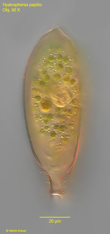
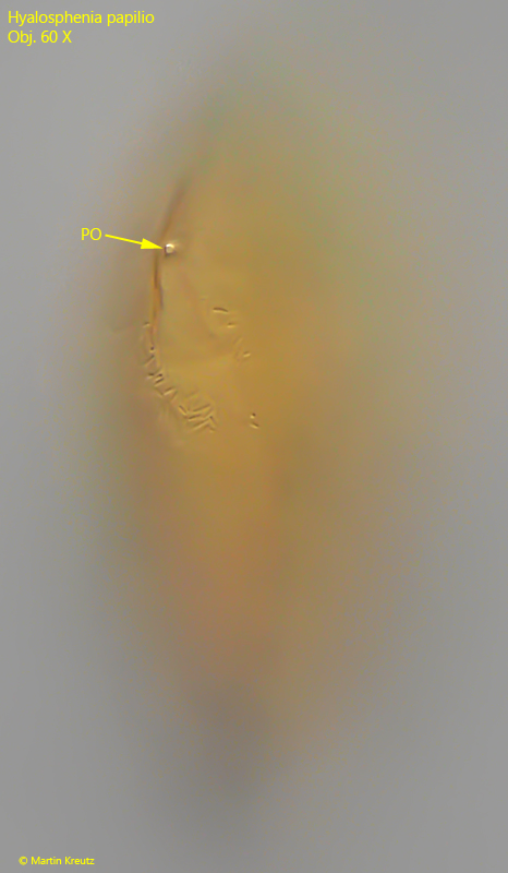
The test is strongly flattened. In order to obtain a lateral view, the coverslip must be moved carefully when the layer thickness is high. In favorable cases, the lateral pores can then also be seen (s. fig. 6).
Fig. 1 a-b:Hyalosphenia papilio. L = 133 µm (of test). Two focal planes of an unsquashed specimen. Obj. 40 X.
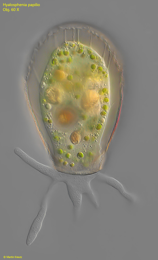
Fig. 2:Hyalosphenia papilio. L = 125 µm (of test). An elongated specimen with several pseudopodia. Obj. 60 X.
Fig. 3:Hyalosphenia papilio. L = 116 µm (of test). Lateral view of the compressed test. Obj. 60 X.
Fig. 4:Hyalosphenia papilio. L = 125 µm (of test). Focal plane on the contractile vacuoles (arrows) located in the posterior half of the protoplast. Obj. 60 X.
Fig. 5:Hyalosphenia papilio. L = 128 µm (of test). An empty test with the two lateral pores (PO). Obj. 40 X.
Fig. 6:Hyalosphenia papilio. Lateral view of a specimen with focal plane on one of the two the lateral pores (PO) in the posterior third of the test. Obj. 60 X.