head trilobed, elongated oval, almost wide as body
neck region distinctly constricted
pharynx elongated cylindrical
anterior end of intensine with a brownish ring
dorsal cuticle thin, flexible, scales absent
ventral side naked, scales absent
furca 17–18 µm long
toes conical shaped
adhesive tubes 4–5 µm long
Ichthydium bifasciale
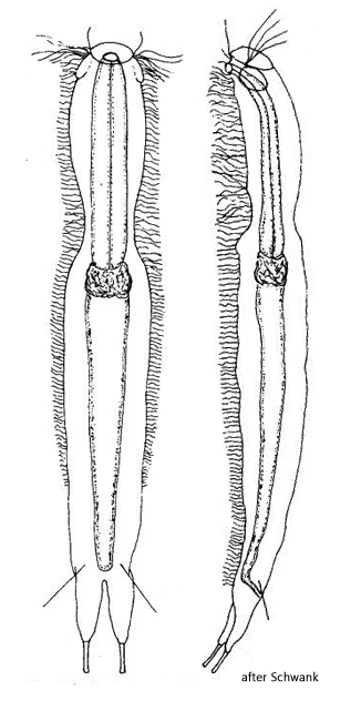
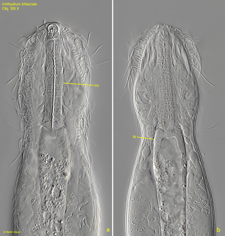
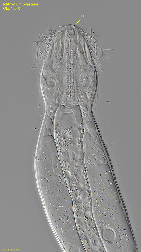
In December 2022, I found a 162 µm long gastrotrich in the mud of the Simmelried, with features suggesting an assignment as Ichthydium bifasciale. The head is long oval and slightly narrowed towards the front. Apically, there is an almost rectangular cephalion that slightly protrudes (s. fig. 5). According to Schwank (1990), the soft cuticle of Ichthydium bifasciale is completely naked. Only a few species within the genus Ichthydium have a slight longitudinal striping on the dorsal side (e.g. Ichthydium fossae). However, in my specimen, I was able to recognize very delicate scales at high magnification, both on the dorsal and ventral sides (s. figs. 6 and 8). These very delicate scales are lanceolate, with a distal, simple spine (s. fig. 7). The scales are 12–15 µm long. The spines account for about 60% of the scale length. The spines overlap the lanceolate parts of the scales, giving the impression that they are keeled. However, this is not the case. Identical scales are found on the ventral side (s. fig. 9). The toes are very short, about 10 µm, and conically shaped (s. fig. 10). At the entrance of the intestine, a ring-shaped thickening can be seen, which is typical for the genus Ichthydium (s. fig. 4 b). The pharynx is almost cylindrical, without thickening, and about 40 µm long (s. fig. 4 a).
Although my specimen is about 15% longer than described by Schwank and delicate scales could be observed, it still appears to be Ichthydium bifasciale, as both the typical body shape with the elongated head and the shape of the intestine, pharynx, and furca match. My specimen is also identical to a specimen of Ichthydium bifasciale found and photographed by Dr. Michael Müller in 2019 (s. Mikroskopie Forum – Ichthydium bifasciale). However, he was unable to detect scales on his specimen, which may have been due to the fact that the images were taken with oblique lighting, which could not contrast the very delicate scales. Verification of whether the scales of Ichthydium bifasciale are a constant feature can only be verified with further findings.
The similar species Ichthydium brachykolon does not have a rectangular cephalion, the toes are significantly longer (17–18 µm), and the head is wider than the body. Additionally, the pharynx of Ichthydium brachykolon has a distinct thickening in the middle, and the body is somewhat smaller at about 130 µm.
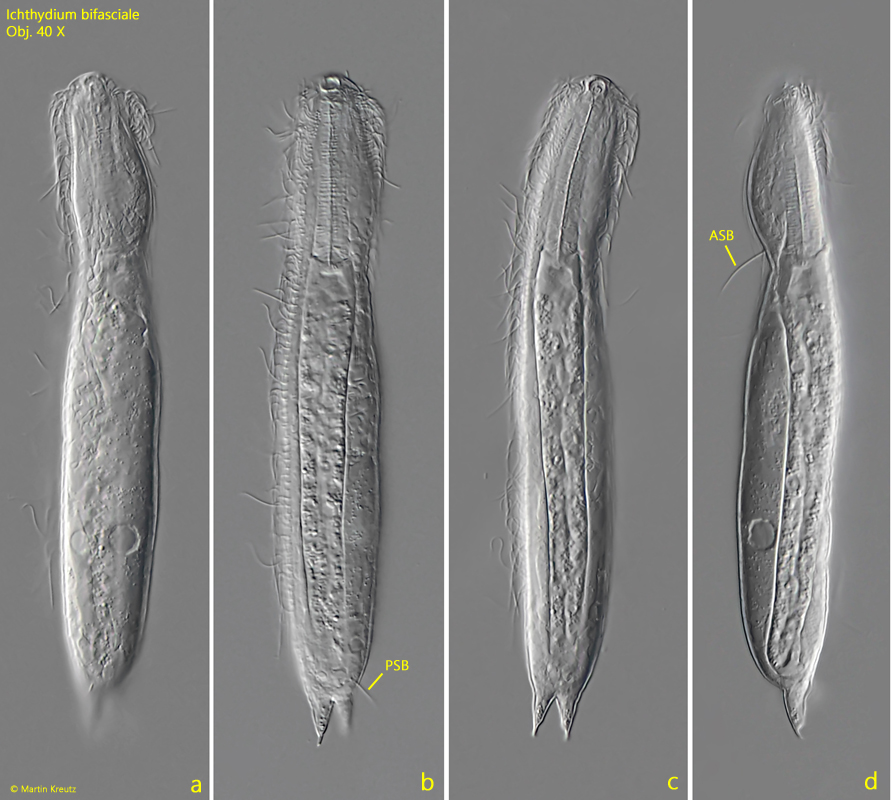
Fig. 1 a-d:Ichthydium bifasciale. L = 162 µm. A freely swimming specimen from dorsal (a–c) and from right (d). ASB = anterior sensory bristle, PSB = posterior sensory bristle. Obj. 40 X.
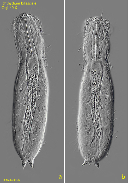
Fig. 2 a-b:Ichthydium bifasciale. L = 162 µm. The slightly squashed specimen as shown in fig. 1 a-d. Obj. 40 X.
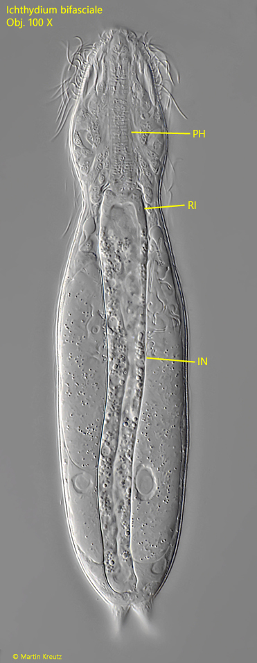
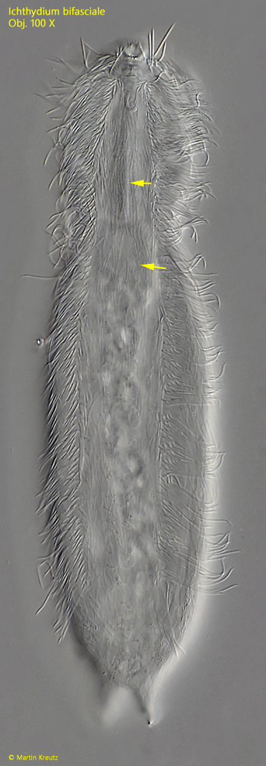
Fig. 3:Ichthydium bifasciale. L = 162 µm. Total view from dorsal of the slightly squashed specimen as shown in fig. 1 a-d. IN = intestine, PH = pharynx, RI = ring-shaped thickening of the intestine entrance. Obj. 100 X.
Fig. 4 a-b:Ichthydium bifasciale. Two focal planes of the head. PH = pharynx, RI = ring-shaped thickening of the intestine entrance. Obj. 100 X.
Fig. 5:Ichthydium bifasciale. Focal plane on the almost rectangular shaped kephalion (KE). Obj. 100 X.
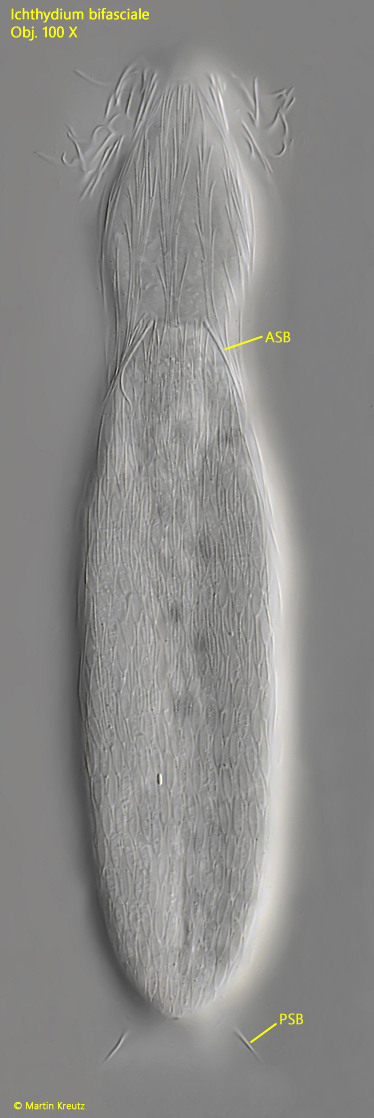
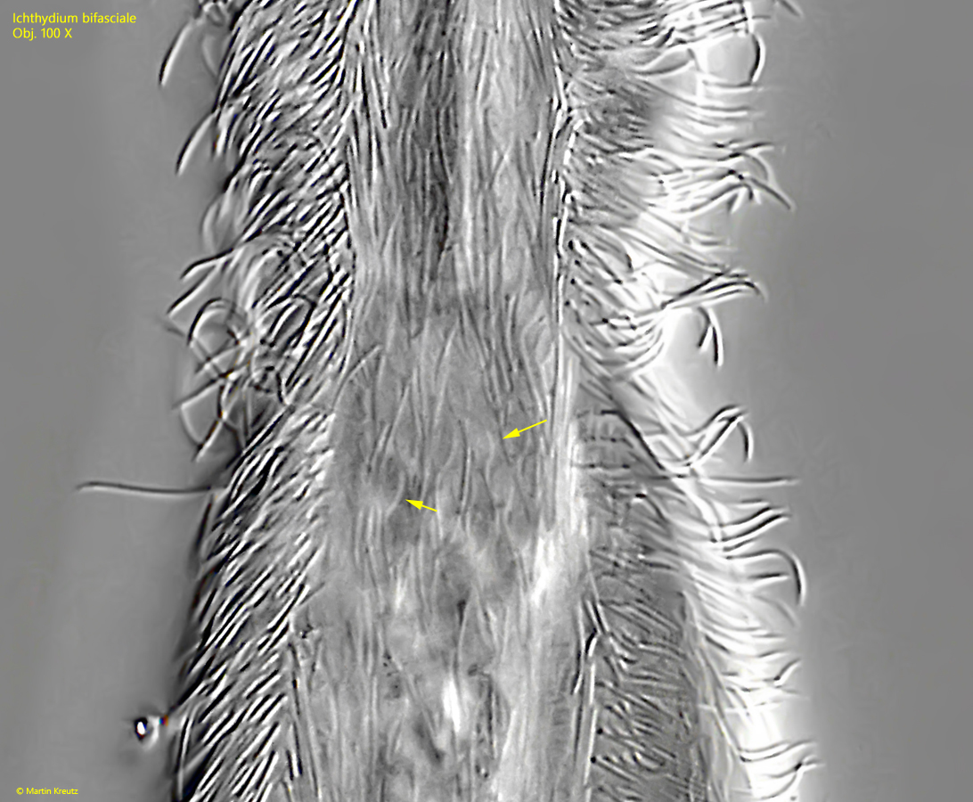
Fig. 6:Ichthydium bifasciale. Focal plane on the delicate, lanceolate shaped scales of the dorsal side. ASB = anterior sensory bristle, PSB = posterior sensory bristle. Obj. 100 X.
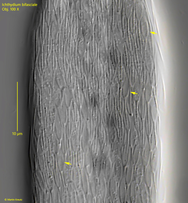
Fig. 7:Ichthydium bifasciale. The lanceolate shaped scales of the dorsal side in detail (arrows). Each scale has a simple spine at the distal end with a length of 5–7 µm. The lanceolate shaped part of the scales has a length of 6–7 µm. Obj. 100 X.
Fig. 8:Ichthydium bifasciale. Focal plane on the delicate, lanceolate shaped scales of the ventral side. Obj. 100 X.
Fig. 9:Ichthydium bifasciale. A strongly contrasted part of the image as shown in fig. 8 for visualisation of the ventral scales. They are from the same type as the dorsal scales. Obj. 100 X.
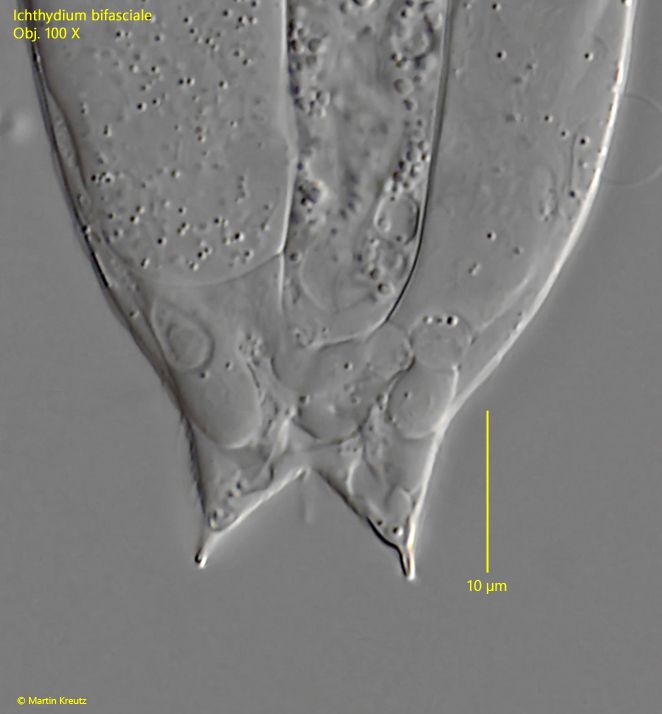
Fig. 10:Ichthydium bifasciale. The short, conical shaped toes with a length of about 10 µm in a squashed specimen. Obj. 100 X.