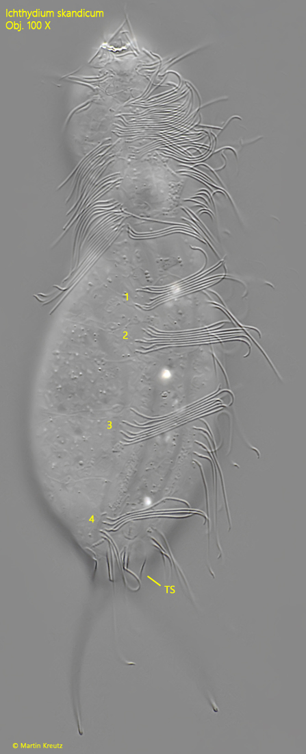
ventral ciliation bands separated in 4 pairs of tufts
ventral two subrectangular terminal scales
Ichthydium skandicum
I have found three specimens of Ichthydium scandicum so far. Two specimens came from the Simmelried and one from the effluent of the seawage plant Constance. The latter location is quite surprising, as the effluent is heavily eutrophicated.
Ichthydium skandicum was only described by Kanneby et al. in 2009. This may be due to the fact that the features for identification are not easy to see. The main feature are the 4 pairs of scales, which are located dorsally directly at the base of the furcae (s. fig. 3). Apart from the scales from which the sensory bristles arise (s. fig. 3), the entire dorsal side is naked. This arrangement of the dorsal scales can only be recognized in strongly squashed specimens. Otherwise Ichthydium skandicum is very similar to the similar species Ichthydium forficula and Ichthydium tanytrichum, which, however, are naked on the dorsal side. In addition, Ichthydium forficula is somewhat larger and more slender while Ichthydium tanytrichum is smaller and stockier.
On the ventral side Ichthydium scandicum is naked apart from the two almost rectangular terminal scales (s. figs. 4 b and 5). The two ventral ciliary bands are not continuous but divided into 4 separate tufts (s. fig. 5).
The specimens of my population were 135–136 µm long. Obviously, the range of variation is very small and corresponds exactly to the range given by Kanneby et al. (132–142 µm).
Fig. 1 a-b:Ichthydium skandicum. L = 136 µm. Two focal planes of a slightly squashed specimen from dorsal. Note the two posterior sensory bristles (PSB) on papillae. The pharynx (PH) is slightly swollen at both ends. ASB = anterior sensory bristles, AT = adhesive tubes. Obj. 100 X.
Fig. 2 a-b:Ichthydium skandicum. Two focal planes of the posterior end from dorsal with the pincer-shaped adhesive tubes (AT). Note the scales (SC) adhering to the base of the tubes and the terminal invagination (TI) between the adhesive tubes. Obj. 100 X.
Fig. 3:Ichthydium skandicum. The arrangement of the dorsal scales (SC) near the posterior end in a squashed specimen. Note the scale of the sensory bristle (SSB). Obj. 100 X.
Fig. 4 a-b:Ichthydium skandicum. L = 135 µm. A second specimen from ventral. Note the almost rectangular terminal scales (TS). Obj. 100 X.
Fig. 5:Ichthydium skandicum. L = 135 µm. The squashed specimen as shown in fig. 3 a-b from ventral. The ventral ciliary bands are separated in 4 tufts (1–4). TS = terminal scales. Obj. 100 X.