cytopharynx armed with long, rod-shaped extrusomes
length about 75 µm
one terminal CV
two spherical macronuclei with each one adjacent micronucleus
cortex with mucilaginous layer
a conspicuous flagellum-like, retractable mouth process
Ileonema ciliata
I have only found Ileonema ciliata once so far in March 2024 in a sample of mud from the Simmelried. On this occasion I was able to observe about 10 specimens.
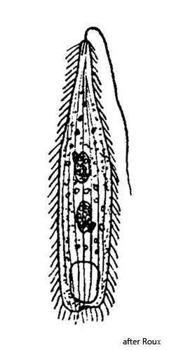
Ileonema ciliata was first described by Roux (as Monomastix ciliatus). After that there are obviously no further documented finds. Kahl (1930) obviously adopted Roux’s drawing and description. It is not clear from his brief description that he found and examined specimens himself.
The specimens of Ileonema ciliata that I examined deviate from Roux’s description, particularly with regard to body length. My specimens were 120–145 µm long (elongated specimens, without mouth flagellum), whereas Roux gives a length of only 75 µm. Therefore, my specimens would rather fit the description of Ileonema chobicola (Foissner, 2016). This species also has two macronuclei and two micronuclei, but is 120–225 µm long. The structure of the dorsal brush with pairs of club-shaped, short bristles (s. fig. 5 a-b) also corresponds to the shape of the dorsal brush of Ileonema chobicola, as described by Foissner. However, the shape of the extrusomes of the specimens in my population differs from those of Ileonema chobicola. My specimens had slightly curved, thin rods with a length of 19–22 µm. The rods were consistently thick, without any thickening (s. fig. 7). In contrast, the extrusomes of Ileonema chobicola are just as long (18–22 µm) but needle-shaped, with a clearly thickened end. I could not observe this shape in my specimens. In addition, Ileonema chobicola was isolated by Foissner from the dry sediment of a river (Chobe River) in Botswana. Thus, the nature of the localities differ considerably.
Since only the few data on Ileonema ciliata by Roux are available so far and because it is no longer possible to reconstruct how Roux carried out the length measurement of his specimens (possibly on contracted specimens), I would like to stick to the identification of my specimens as Ileonema ciliata, especially because of the deviation from the shape of the extrusomes as described for Ileonema chobicola and the different nature of the sampling sites.
The two species Ileonema ciliata and Ileonema chobicola differ only in very few features (length, extrusomes). Further studies may show that the two species are synonymous in terms of variability. However, this would require more findings and data, especially from Ileonema ciliata, in order to check the constancy of the characteristics.
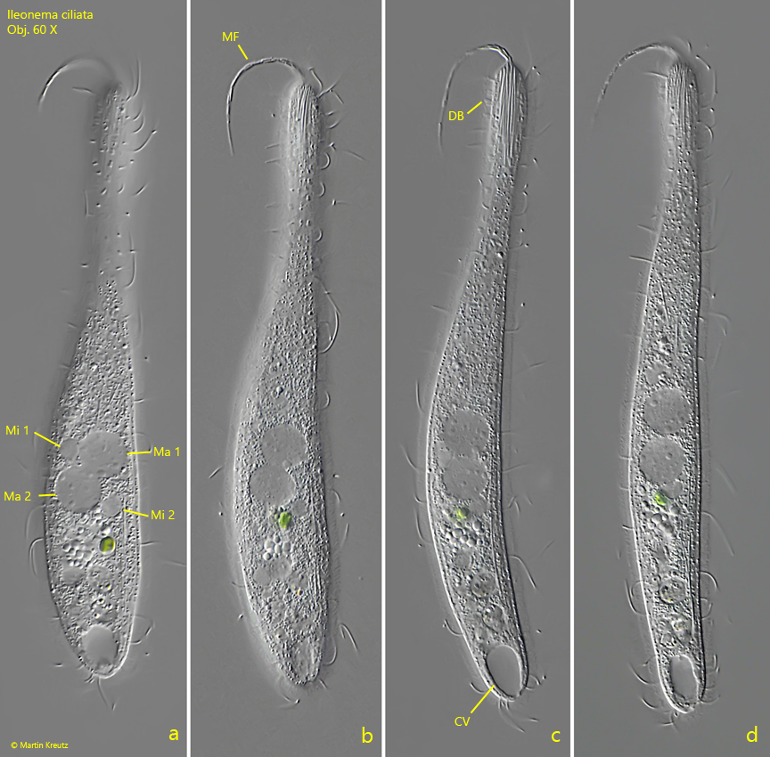
Fig. 1 a-d:Ileonema ciliata. L = 128 µm. A freely swimming specimen with the characteristic mouth flagellum (MF). CV = contractile vacuole, DB = dorsal brush, Ma 1+2 = macronuclei, Mi 1+2 = micronuclei. Obj. 60 X.
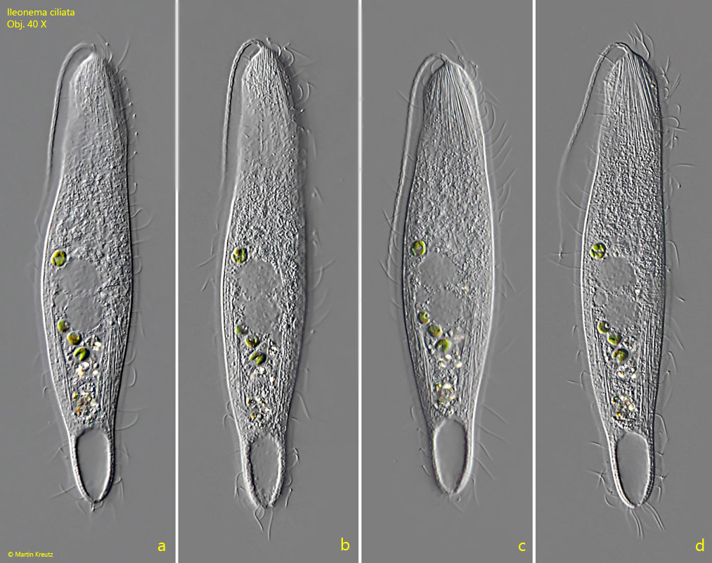
Fig. 2 a-d:Ileonema ciliata. L = 140 µm. A second specimen with a 63 µm long mouth flagellum. Obj. 40 X.
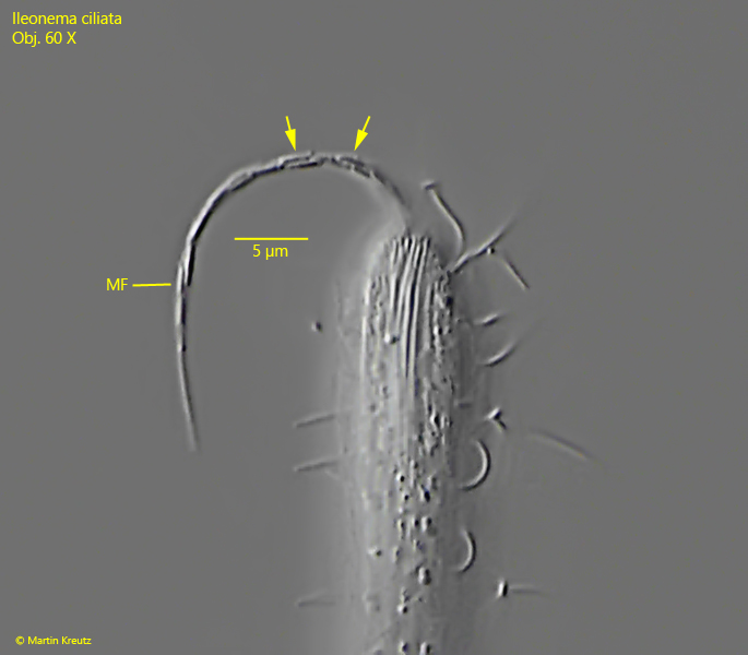
Fig. 3:Ileonema ciliata. The mouth flagellum in detail. It contains short rods with a length of 2.0–2.3 µm (arrows). MF = mouth flagellum. Obj. 60 X.
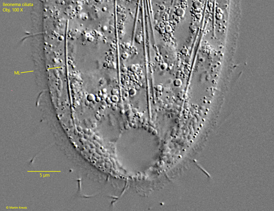
Fig. 4:Ileonema ciliata. A squashed specimen with focal plane on the base of the mouth flagellum, where two short rods are visible (RMF). The body is covered by mucilaginous layer (ML) with a thickness of 2–2.5 µm. Ma 1+2 = macronuclei, Mi = one of the two micronuclei. Obj. 100 X.
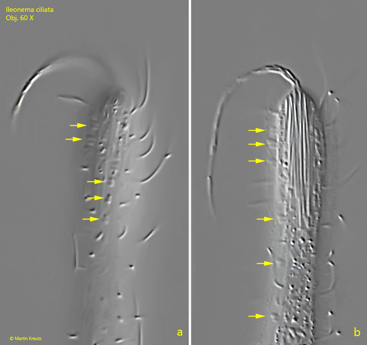
Fig. 5 a-b:Ileonema ciliata. Two focal planes on the dorsal brush with characteristic pairs of short, club-shaped bristles (arrows). Obj. 60 X.
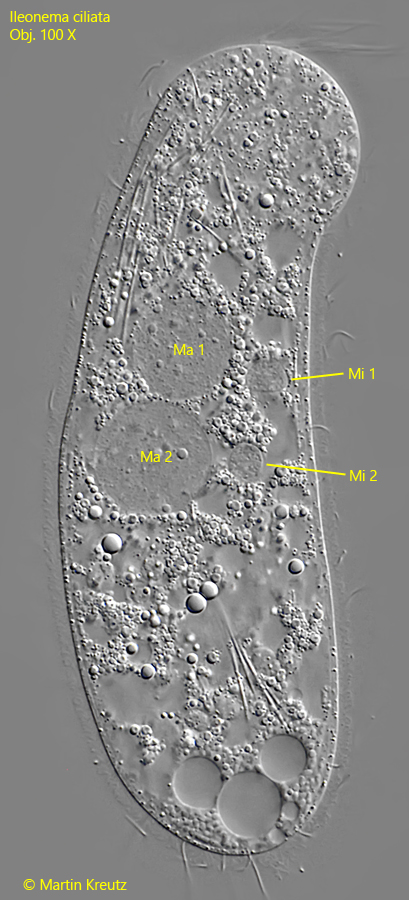
Fig. 6:Ileonema ciliata. A squashed specimen with focal plane on the two macronuclei (Ma 1, Ma 2) and the two adjacent microcuclei (Mi 1, Mi 2). Obj. 100 X.
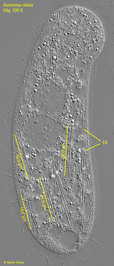
Fig. 7:Ileonema ciliata. The same specimen as shown in fig. 6 with focal plane on the rod-shaped, slightly curved extrusomes (EX) with a length of 19–22 µm. Obj. 100 X.
Fig. 8:Ileonema ciliata. At the limit of the possible resolution the mucilaginous layer (ML) revealed a granulated structure. This is caused by lepidosomes scattered in the gelatinous mass. The complex structure of the lepidosomes is below the resolution of a light microscope. Obj. 100 X.