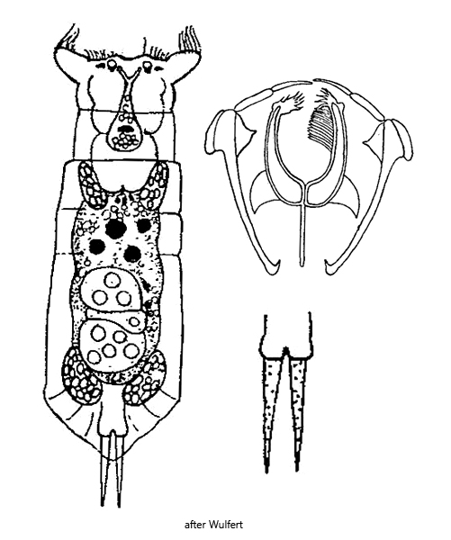
stomach and blind sacs green due to kleptoplasts from prey
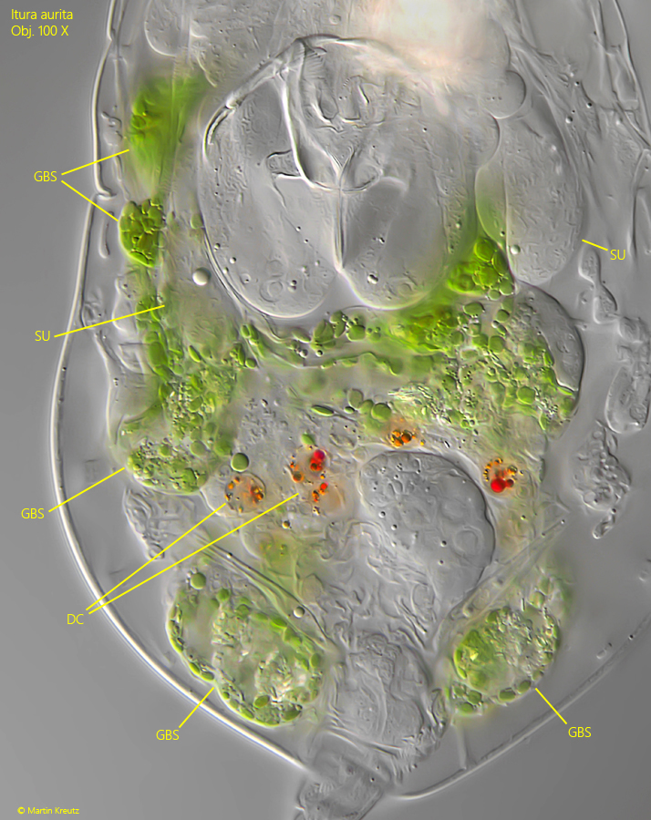
gastric glands absent
two elongated subcerebral glands present
reddish, orange or brown defecation concretions
food with three pseudosegments
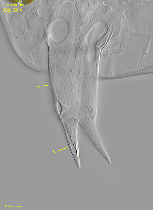
toes straight, slender and pointed
feeding on euglenids
Itura viridis
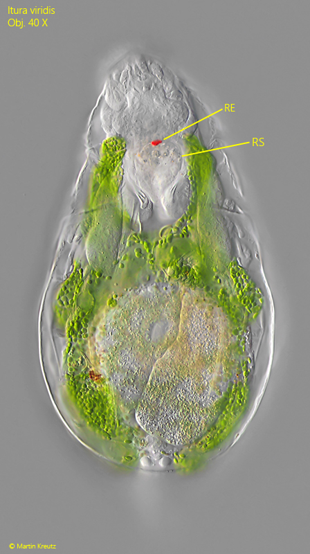
I mainly find Itura viridis among floating and decomposing plants. When examining the samples, the specimens are easy to recognize by their size and bright green color. Only free-swimming specimens extend their lateral auricles at the head (s. figs 1 a and 2 a).
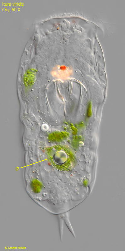
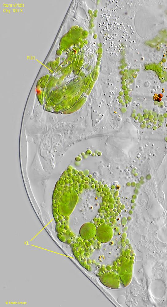
Itura viridis is one of the few rotifers that possess symbiotic zoochlorellae. These are located in the blind sacs of the stomach. The blind sacs are bladder- and finger-shaped protrusions of the stomach, which extend high up to the mastax and into the posterior abdomen (s. fig. 11). The zoochlorellae are chloroplasts that essentially originate from phagocytized euglenids such as Euglena or Phacus. One should therefore rather speak of kleptoplasts here, since they were “stolen” from the prey. In the gastric blind sacs, the kleptoplasts continue to carry out their synthetic activity and additionally supply Itura viridis with nutrients.
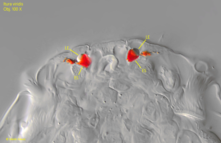
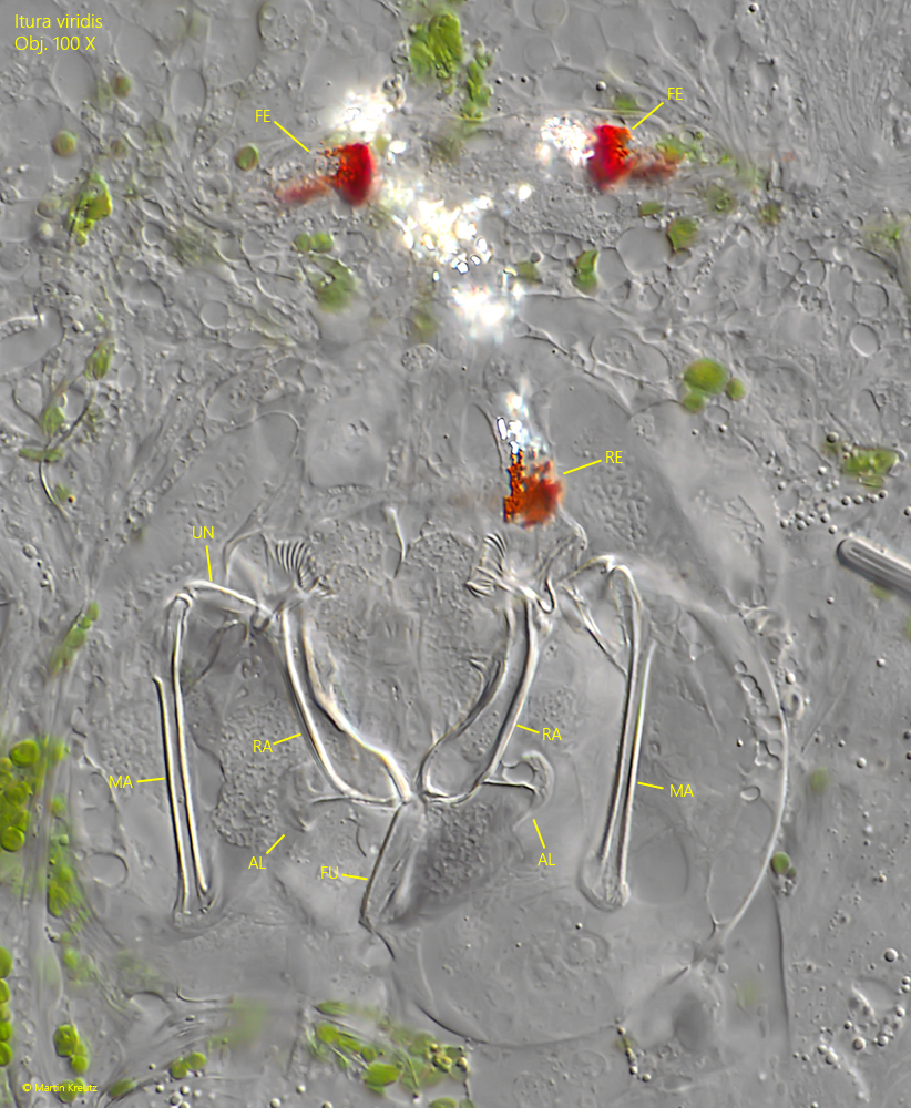
Itura viridis has no gastric glands. Instead, there are two subcerebral glands at the level of the mastax (s. fig. 11). At the front end of the head there are two eyespots with lenses (s. fig. 7). A third eyespot is located on the retrocerebral sac (s. fig. 5). This is usually filled with highly refractive crystals (s. fig. 10). In the center of the stomach, so-called defecation concretions are visible (s. fig. 11). These are orange, red or brown residues of the digestion process that have an oily character.
The specimens of Itura viridis in my population were always 190–240 µm long. I have never found larger specimens. This size is clearly below the range of 260–400 µm given by Koste (1978). All other characteristics agree with the description of Itura viridis.
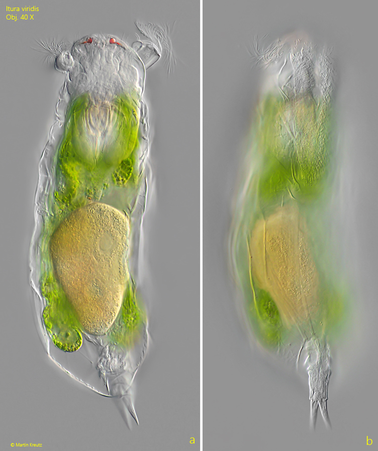
Fig. 1 a-b: Itura viridis. L = 210 µm. A freely swimming specimen from dorsal (a) and from right (b). Obj. 40 X.
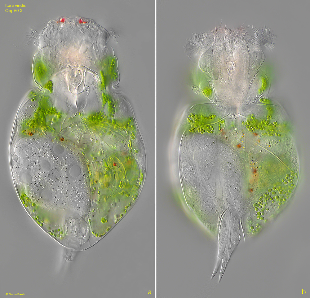
Fig. 2 a-b: Itura viridis. L = 190 µm. A second freely swimming specimen from dorsal (a) and from ventral (b). This specimen carries an orange-colored egg. Obj. 60 X.
Fig. 3 a-b: Itura viridis. L = 190 µm. The slightly squashed specimen as shown in fig. 2 a-b from dorsal (a) and from ventral (b). Obj. 60 X.
Fig. 4 a-b: Itura viridis. L = 230 µm. A third freely swimming specimen from dorsal (a) and from ventral (b). Obj. 60 X.
Fig. 5: Itura viridis. A slightly squashed specimen from dorsal with focal plane on the retrocerebral eyespot (RE) and on the retrocerebral sac (RC). Obj. 40 X.
Fig. 6: Itura viridis. A specimen with an ingested Phacus spec. (IP). Obj. 60 X.
Fig. 7: Itura viridis. The two frontal eyesspots (ES) with lenses (LE). Obj. 100 X.
Fig. 8: Itura viridis. The frontal eyespots (FE), the retrocerebral eyespot (RE) and the trophi below in a strongly squashed specimen. AL = alulae, FU = fulcrum, Ma = manubria, RA = rami, UN unci. Obj. 100 X.
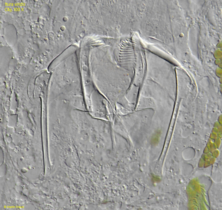
Fig. 9: Itura viridis. The trophi in a second strongly squashed specimen. Obj. 100 X.
Fig. 10: Itura viridis. The retrocerebral sac (RCS) is filled with highly refractive crystals. The short excretory duct (ED) is also visible. FE = frontal eyespots. Obj. 100 X.
Fig. 11: Itura viridis. The gastric blind sacs (GBS) are mainly filled with cleptoplasts taken from ingested euglenids. In the center orange colored defecation concretions are visible. Beside the mastax the two elongated subcerebral glands (SU) are visible. Obj. 100 X.
Fig. 12: Itura viridis. Two gastric blind sacs filled with kleptoplasts (KL). The kleptoplasts are chloroplasts taken from ingested euglenids. In the upper gastric blind sac the residues of an ingested Phacus spec. (PHR) are visible. Obj. 100 X.
Fig. 13: Itura viridis. The foot (FT) with the two toes (TO) in detail. Obj. 100 X.