often spherule of polyphosphate and cyanophycine in center of cells
sometimes with refractive granules near crosswalls
benthic lifestyle
Kamptonema chlorinum
Kamptonema chlorinum was originally described by Kützing (1892) as Oscillatoria chlorina. Other authors described the species as Phormidium chlorinum or Lyngbya chlorina. In 2014, Strunecký, Komárek & J. Smarda transferred the species to the new genus Kamptonema based on morphological and genetic analyses and named it Kamptonema chlorinum.
So far I have only found the cyanobacterium Kamptonema chlorinum in the Simmelried, where it is extremely common. It lives on the surface of the mud layer and forms large mats. In old samples it grows up the walls of the sample container.
Kamptonema chlorina is strongly birefringent. This means that its “true” color (yellowish-green) can only be observed in bright field (s. figs. 1 and 2). In DIC and also between crossed polarization filters, its color changes very strongly from green to orange to violet (s. figs. 5 a-b and 6 a-b) . The filaments also light up strongly. It is not known which structures are responsible for the birefringent properties of the filaments.
Other characteristic features of Kamptonema chlorinum are a hyaline cap on the terminal cells (s. figs. 2 and 3 a-b) and a clear, narrow transverse striation of the cells (s. fig. 4 a-b). These transverse striation is caused by cross wall which that are not completely closed, similar to an aperture diaphragm. This leaves a central opening in the middle. These pseudo-crosswalls are arranged in a distance of approx. 0.5 µm. Adjacent to the “real” cross walls, which separate the cells, there are often few, highly refractive grains (s. fig. 4 b).
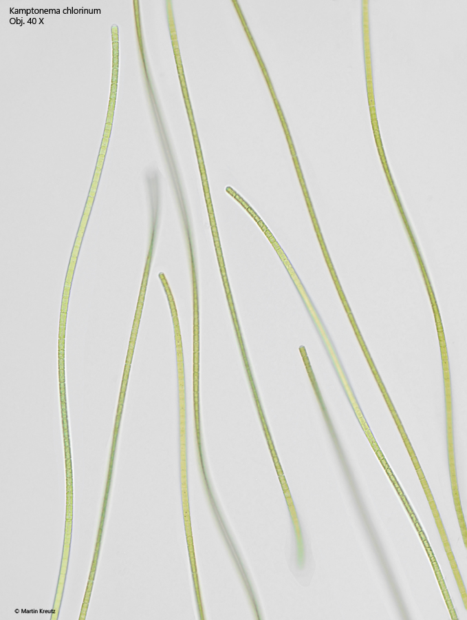
Fig. 1:Kamptonema chlorinum. Several trichomes in brightfield illumination. Obj. 40 X.
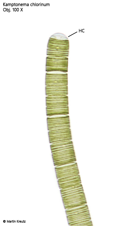
Fig. 2:Kamptonema chlorinum. D = 4.2 µm. In brightfield illumination the color of the filaments is yellowish-green. Note the hyaline cap of the terminal cell (HC) and the tight transverse striation. Obj. 100 X.
Fig. 3 a-b:Kamptonema chlorinum. D = 4.4 µm. Two focal planes of the terminal cell with a hyaline cap (HC) in DIC. Obj. 100 X.
Fig. 4 a-b:Kamptonema chlorinum. D = 6.8 µm. Two focal planes of a filament. The cross walls (CW) can be continuous or, similar to an aperture diaphragm, still open in the middle. The distance between the cross walls and the pseudo-cross walls is about 0.5 µm. Near the cross, refractive granules (RG) can be found. Obj. 100 X.
Fig. 5 a-b:Kamptonema chlorinum. The filaments are birefringent, which leads to different colors in the quadrants in the DIC (a) and to a clear refraction cross between crossed polarization filters (b). Obj. 20 X.
Fig. 6 a-b:Kamptonema chlorinum. A short filament shows different colors in DIC depending on the angle between the orientation of the filament to the angle of the used polarized ligth. Obj. 100 X.