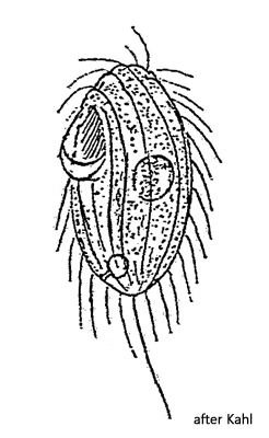
contractile vacuole subterminal with short emptying canal
macronucleus globular, mid-body
one spherical micronucleus
sometimes extrusomes present
one caudal cilium
Zitheron muscorum
Zitheron muscorum was first described by Kahl (1931) as Saprophilus muscorum. Corliss (1960) transferred the species to the genus Sathrophilus, and Jankowski (2007) finally transferred it to the newly created genus Kariphilus. Finally, Kariphilus muscorum was trasnferred by Poláková et al. (2021) to the new genus Zitheron. The genus Zitheron was also raised by Jankowski in 2007.
I found Zitheron muscorum in a moss sample in March 2021. Kahl also found the population he describes in a few lines in moss. The ciliate has a relatively large mouth opening compared to its body size, which is located in the anterior half. A membranelle of long cilia is visible inside. At 25–28 µm, my specimens were smaller than those described by Kahl (35 µm).
Apart from this deviation, all other characteristics correspond to Kahl’s description. Particularly striking and explicitly mentioned by Kahl is the short emptying canal of the subterminal contractile vacuole (s. fig. 1 c). In my specimens, I was able to identify isolated, rod-shaped extrusomes with a length of 1.8 µm (s. fig. 1 f). In the cytoplasm of various specimens, I was able to identify symbiotic bacteria, but these were difficult to contrast. The margin of the ciliate appears slightly wavy due to indentations where the cilia originate.
Fig. 1 a-f:Zitheron muscorum. L = 26 µm. Different focal planes of a freely swimming specimen. Note the short emptying canal (EC) of the subterminal contractile vacuole (CV). CC = caudal cilium, EX = extrusome, Ma = macronucleus, Mi = micronucleus. Obj. 100 X.