dorsal with 3 median fields, first field square like
lorica with fine reticulate pattern
length 110–150 µm
ventral side flat
dorsal side convex
two long anterior median spines
posterior spines absent
one large eyespot
Keratella ticinensis
I find Keratella ticinensis very frequently in the Simmelried. The species is permanently present there, especially in floating plant masses.
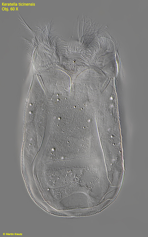
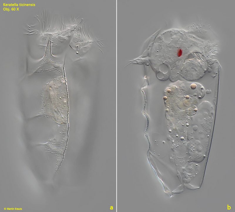
Keratella ticinensis can easily be recognized by the shape of the lorica. It only has spines on the anterior margin. The lorica is broadly rounded at the posterior end, without any spines. In addition, the lorica has a strikingly fielded dorsal side. On this is a median row of 3 fields, the first of which is square-shaped. The other two fields are hexagonal. Another important feature is the very large eyespot, which can be clearly seen even at low magnifications.
Fig. 1 a-d:Keratella ticinensis. L = 135 µm. Different focal planes of a slightly squashed specimen from dorsal. Note the 3 median fields of the lorica (1–3) and the two anterior median spines (AMS). ES = eyespot, St = stomach. Obj. 60 X.
Fig. 2:Keratella ticinensis. The dorsal side of a squashed specimen. Note the fine reticulate pattern and the three median fields (1–3). The first field is square shaped, while the fields 2 and 3 are hexagonal. Obj. 100 X.
Fig. 3:Keratella ticinensis. L = 129 µm. The ventral side is smooth without hexagonal fields. Obj. 60 X.
Fig. 4 a-b:Keratella ticinensis. L = 129 µm. Two focal planes from right. Obj. 60 X.
Fig. 5:Keratella ticinensis. The trophi in a strongly squashed specimen. Obj. 100 X.