So far I have only found a few specimens of Kirchneriella irregularis. All specimens came from the uppermost mud layer in the Simmelried.
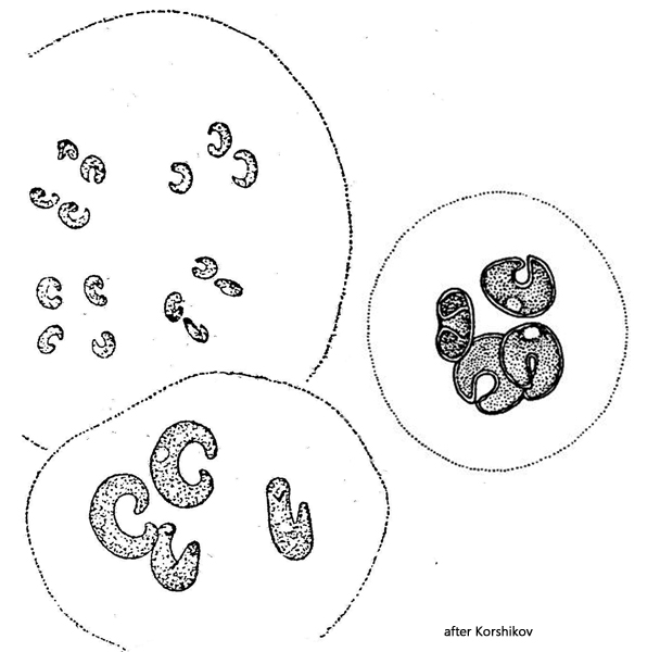
The cells of Kirchneriella irregularis are strongly curved and at the same time somewhat twisted. As a result, the apices do not meet, but overlap slightly (s. fig. 4). In frontal view the cells appear almost circular. In the literature (Huber-Pestalozzi, 1983) the presence of a pyrenoid is considered possible, but I was unable to detect one in any of the specimens in my population. Reproduction takes place by autospores with 4 daughter cells each (s. fig. 2 a-b). The length of the adult cells in my population was between 10.0–11.5 µm. The colonies were always very small. Mostly less than 8 cells, which lay in a very delicate, barely perceptible mucus envelope.
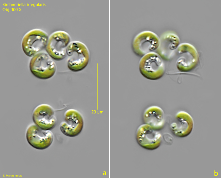
Fig. 1 a-b:Kirchneriella irregularis. L = 7.5–8.8 µm. Two focal planes of a small colony of 7 cells. Obj. 100 X.
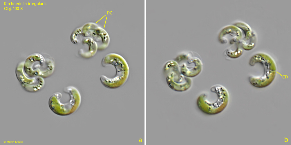
Fig. 2 a-b:Kirchneriella irregularis. L = 10.1-11.2 µm. A second small colony with two autospores with 4 daugther cells (DC) each. In one of the parent cells a cell division (CD) starts. Obj. 100 X.
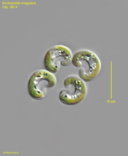
Fig. 3:Kirchneriella irregularis. L = 5.0–5.2 µm. Four young daughter cells. Obj. 100 X.
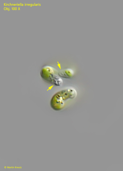
Fig. 4:Kirchneriella irregularis. Two cells that are perpendicular to the focal plane. As the cells are slightly twisted, the two apices overlap slightly (arrows). Obj. 100 X.