striation of pellicle run almost parallel to body axis
body not metabol, rarely U- or S-shaped
Lepocinclis acus
Some species of the genus Euglena were transferred to the genus Lepocinclis by Marin and Melkonian in 2003, as the body of these species is rigid and rotund. This also included Euglena acus.
Lepocinclis acus is one of the most common euglenids in my samples. I find it in almost all my localities. This species can even be found in plankton samples.
Lepocinclis acus is easily recognized by its spindle or almost needle-like shape. The body is not metabolic. Only rarely do the cells bend in a U- or S-shape. They swim in a straight line without changing direction much. The numerous chloroplasts are arranged parietally, i.e. close to the cell wall (s. figs. 2 b and 3 b). They almost always contain large, oblong-shaped paramylon grains (s. figs. 2 a and 3 a). Depending on the nutritional state of the cells, they can be thicker or thinner. The cell nucleus is located almost centrally (s. fig. 2 a). The striation of the pellicle is either parallel to the longitudinal axis (s. fig. 3 c) of the cells or with a slight clockwise rotation (s. fig. 2 c).
Fig. 1 a-c:Lepocinclis acus. L = 190 µm. A freely swimming specimen. Obj. 60 X.
Fig. 2 a-c:Lepocinclis acus. L = 175 µm. The focal planes of a slightly squashed specimen. Note the disc-shaped chloroplasts (Chl) and the olong paramylon rods (PG). ES = eyespot, F = flagellum, Nu = nucleus, RE = reservoir, SP = striation of the pellicle. Obj. 100 X
My population of Lepocinclis acus is very often infested by an endoparasitic fungus (s. fig. 3 a). The motile spores of the fungus invade the cells and form immobile, oblong or oval-shaped stages that grow rapidly and later occupy almost a fourth of host cell. I could not identify this parasitic fungus but I could document some stages of the life cycle (s. figs. 4–8).
Fig. 3 a-c:Lepocinclis acus. The squashed specimen as shown in fig. 1 a-c. Near the posterior end an endoparasitic fungus cell (PC) is visible. The nucleus (Nu) is almost centrally located. Chl = disc-shaped chloroplasts, ES = eyespot, PG = paramylon grains, SP = striation of the pellicle. Obj. 100 X
Fig. 4 a-c:Lepocinclis acus. Three focal planes of a specimen infested by a parasitic fungus cell (PC). NPC = nucleus of the parasitic cell. Obj. 100 X
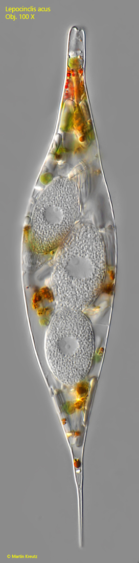
Fig. 5:Lepocinclis acus. A specimen with three parasitic fungus cells. Obj. 100 X
Fig. 6 a-c:Lepocinclis acus. A later stage with a grown parasitic cell (PC). Anterior and posterior to the fungal cell accumulations of hematochrome grains (HG) are visible. The nucleus of the host cell (NHC) is located posterior to the fungal cell. NPC = nucleus of the parasitic fungal cell. Obj. 100 X
Fig. 7 a-b:Lepocinclis acus. At an even later stage, the fungal cell has divided several times and has formed hundreds of motile spores (MSP), which are densely packed in the center of the host cell. HG = hematochrome granules. Obj. 100 X
Fig. 8:Lepocinclis acus. The motile spores (MSP) have been released by the pressure of the coverslip. They are 3–4 µm long and appear to have only one spiralized flagellum. Obj. 100 X