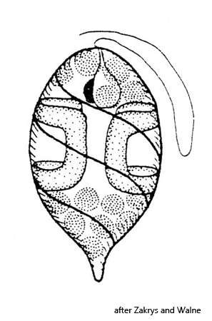
posterior end with a blunt tail, sometimes elongated or spine-shaped
length 13–25 µm
pellicle with a distinct spirally striation, running counterclockwise
one flagellum, about body length
one eyespot in anterior third
two ring-shaped paramylon bodies, arranged laterally
numerous disc-shaped chloroplasts
Lepocinclis ovum
I find Lepocinclis ovum only very rarely in my samples. I may have overlooked specimens due to their small size. In addition, Lepocinclis ovum can be confused with Trachelomonas at small magnifications due to its oval shape.
In my population, the specimens only had a blunt, short spine (s. figs. 1 a and 2 a). However, this can take on very different shapes and can also become much longer or pointed. In addition, my specimens all had a very pronounced and broad striation of the pellicle (s. figs. 1 d and 2 b), which can also be much less pronounced. Therefore, the ring-shaped paramylon grains should also be present for identification (s. fig. 1 c). The body is in apical view rounded (s. fig. 3) and in lateral view mostly oval in shape.
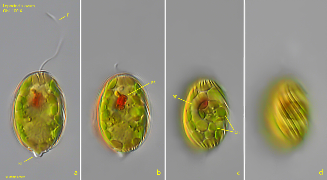
Fig. 1 a-d:Lepocinclis ovum. L = 23 µm. Different focal planes of a slightly squashed specimen. Note the ring-shaped paramylon body (RP) and the distinct striation of the pellicle running counterclockwise to the posterior end. BT = blund tail, Chl = disc-shaped chloroplasts, ES = eyespot, F = flagellum. Obj. 100 X.
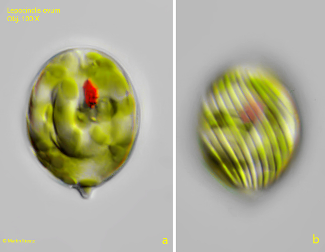
Fig. 2 a-b:Lepocinclis ovum. L = 26 µm. Two focal planes of a second, slightly squashed specimen. Obj. 100 X.
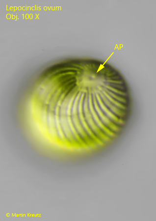
Fig. 3:Lepocinclis ovum. L = 26 µm. The specimen as shown in fig. 2 a-b in apical view. Note the apical porus (AP) where the flagellum arises. Obj. 100 X.