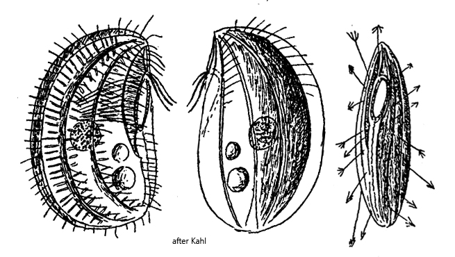
ventral side is more flattened than the convex dorsal side
length 40–50 µm
oral basket broad, clearly visible
macronucleus globular
cytoplasm with green food vacuoles
fusiform extrusomes in furrows of pellicle
two contractile vacuoles below cell equator
feeding on euglenids
Leptopharynx euglenivorus
I found a few specimens of Leptopharynx euglenivorus in May 2026 in samples from the Diebelsee near Johannisthal. The Diebelsee has a moor-like character. The samples were taken from stands of Sphagnum moss mixed with Utricularia spec.
The ciliate Leptopharynx euglenivorus was first described by Kahl in 1926 as “Trichopelma (Leptopharynx) euglenivora”. The name was corrected in 2011 by Foissner, Wolf, Yashchenko & Stoeck to Leptopharynx euglenivorus as an adaptation to the masculine “Leptopharynx”.
Apart from Kahl’s brief descriptions of Leptopharynx euglenivorus, there are no further data or drawings of this ciliate. As important identification features, Kahl lists the clearly visible green food vacuoles and a well-visible and large oral basket. In addition, the ribs on each side of the body are said to meet apically in a short projection (beak-shaped).
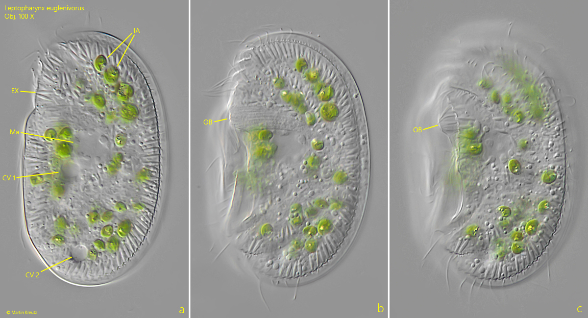
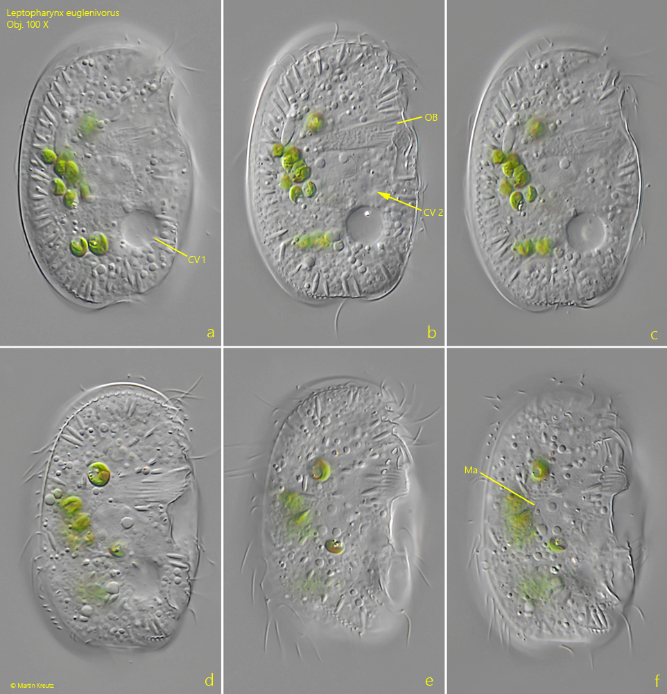
The green food vacuoles were present in all specimens of my population. In Kahl’s population, the green contents came from ingested Lepocinclis spec. (name). In my population, they were very small algae of an unknown genus and source (s. fig. 1 a). But they were not euglenids. I could clearly recognize the oral basket (s. figs. 1 b, 1 c and 2 b) in the anterior third. The lateral ribs were only weakly developed. The extrusomes under the pellicle followed their course. As far as I could see, the ribs also met apically.
The specimens in my population were all between 50–70 µm long and thus larger than described by Kahl (40–50 µm). However, since only a few populations of Leptopharynx euglenivorus have been investigated so far (Kahl found the species only twice), little is known about the variability within the species. Due to the size of the specimens, the algal diet, and the clearly visible oral basket, all characteristics of Leptopharynx euglenivorus are present.
Fig. 1 a-c:Leptopharynx euglenivorus. L = 68 µm. Three focal planes of a slightly squashed specimen from left. Note the two contractile vacuoles (CV 1, CV 2) and the green ingested algae (IA). EX = extrusomes, Ma = macronucleus, OB = oral basket. Obj. 100 X.
Fig. 2 a-f:Leptopharynx euglenivorus. L = 55 µm. A second, smaller specimen from right. CV 1, CV 2 = contractile vacuoles, Ma = macronucleus, OB = oral basket. Obj. 100 X.