In the samples, Linostomella vorticella is easy to recognize by its sack-shaped body with an obliquely truncated front end. The specimens swim quickly and often appear opaque due to the many food vacuoles filling the body. Their preferred food includes Euglena, Chlamydomonas, Chilomonas, Eudorina, and Pandorina.
The mouth opening occupies the entire front half of the body. It is funnel-shaped with a V-shaped, ventral notch. The adoral zone of membranelles is located on the left side and has the shape of a question mark, while the large undulating membrane occupies the right margin of the mouth opening. The terminal contractile vacuole is often difficult to detect due to the mass of food vacuoles in the cytoplasm. The macronucleus is moniliform and consists of 6–10 nodules. In my specimens, there were mostly 6–8 nodules. Even in strongly squashed specimens, I could not detect any micronuclei. Earlier authors also do not mention or describe any. They are either very small or scattered in the cytoplasm and thus difficult to identify there.
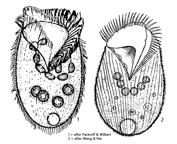
Fig. 1 a-f:Linostomella vorticella. L = 212 µm. A freely swimming specimen from ventral (a, b, d), from left (c, e) and from dorsal (f). Note the adoral zone of membranelles (AZM) on the left side of the oral apparatus and the undulating membrane (UM) on the right side. FV = food vacuoles. Obj. 40 X.
Fig. 2 a-d:Linostomella vorticella. L = 196 µm. A second freely swimming specimen from ventral (a, b, c) and in apical view (d). AZM = adoral zone of membrabelles, OF = oral funnel, UM = undulating membrane. Obj. 40 X.
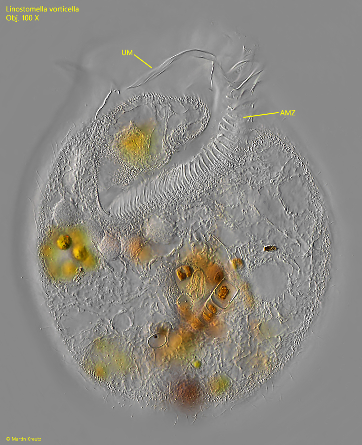
Fig. 3:Linostomella vorticella. A slightly squashed specimen with focal plane on the adoral zone of membranelles (AZM) and the undilating membrane (UM). Obj. 100 X.
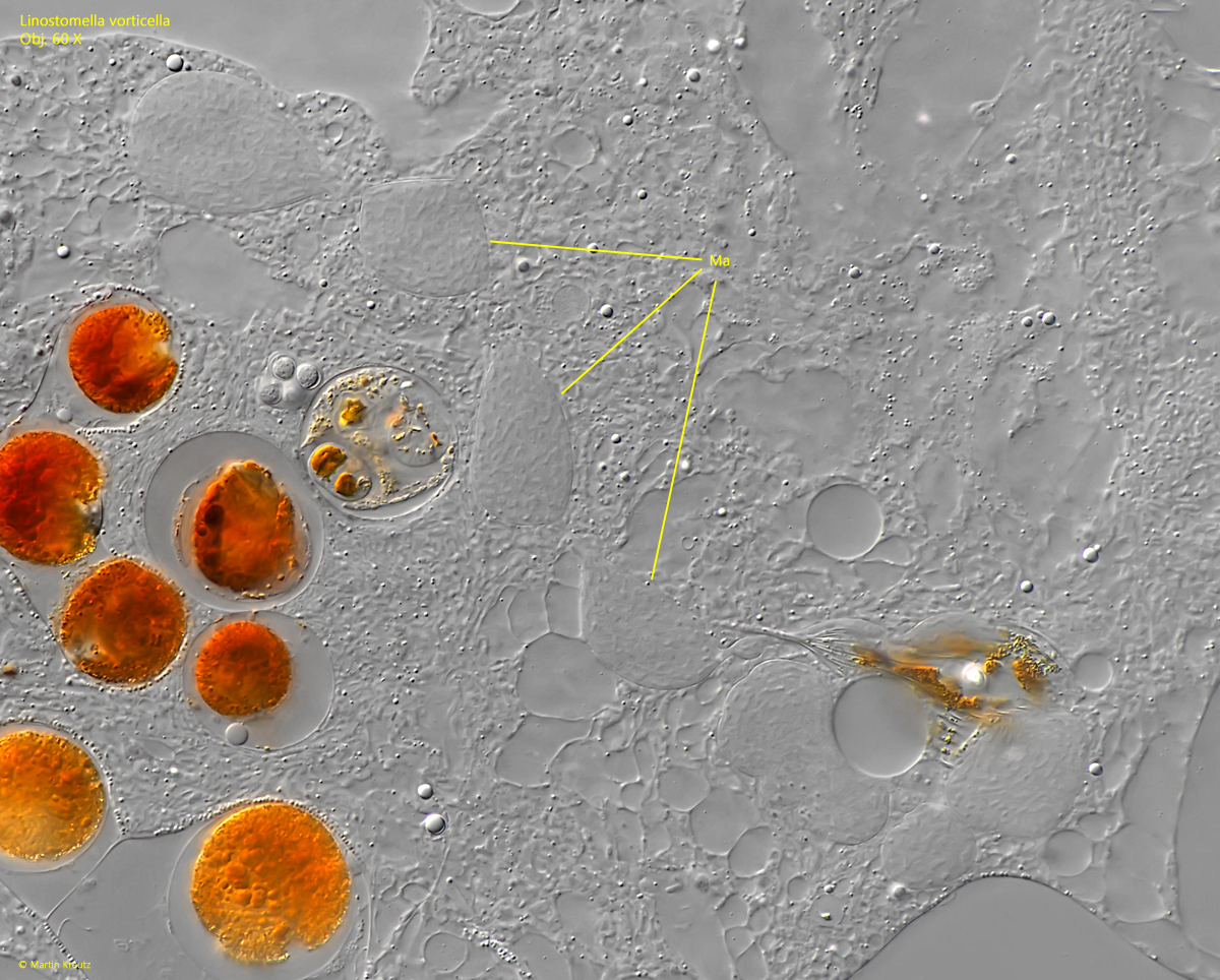
Fig. 4:Linostomella vorticella. The moniliform macronucleus (Ma) of 8 nodules in a squashed specimen. Obj. 60 X.