So far, I have only found Lobomonas ampla in the plankton of the Mühlhalden pond. However, the species occurs there only very rarely. I have only documented two finds in June 1998 and August 2025.
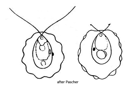
The genus Lobomonas is characterized by a thick gelatinous envelope that is wavy or lobed. The cell has a cup-shaped chloroplast with a pyrenoid, and there is an eyespot in the front third or at the cell equator. The two flagella, which are of equal length, are guided through the gelatinous envelope by thin channels. There are two or more contractile vacuoles at the apical end.
In Lobomonas ampla, the gelatinous sheath is strongly wavy and also encloses the front end of the cell. The gelatinous sheath only fully develops in older cells (Pascher, 1927). Several varieties of Lobomonas ampla have been described, as well as eight species within the genus Lobomonas. Huber-Pestalozzi (1961) assumes that some species could be synonymous, as they only show different stages of development of the gelatinous sheath.
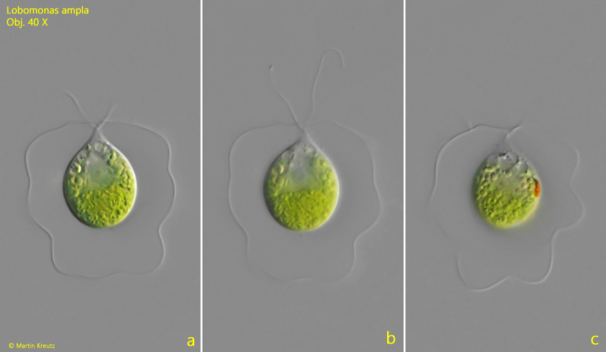
Fig. 1 a-c:Lobomonas ampla. L = 34 µm (of cell). Different focal planes of a freely swimming specimen. Obj. 40 X.
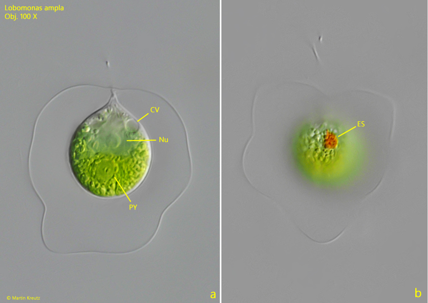
Fig. 2 a-b:Lobomonas ampla. L = 34 µm (of cell). The slightly squashed specimen as shown in fig. 1 a-c. CV = contractile vacuole, ES = eyespot, Nu = nucleus, PY = pyrenoid. Obj. 100 X.
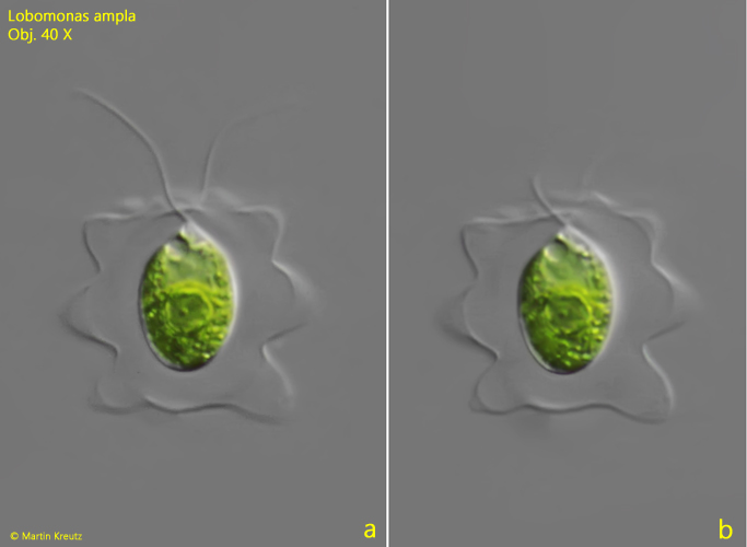
Fig. 3 a-b:Lobomonas ampla. L = 21 µm (of cell). A freely swimming specimen found 1998 in the Mühlhalden pond. Obj. 40 X.