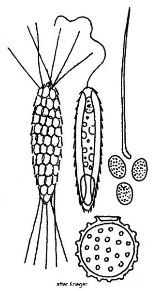
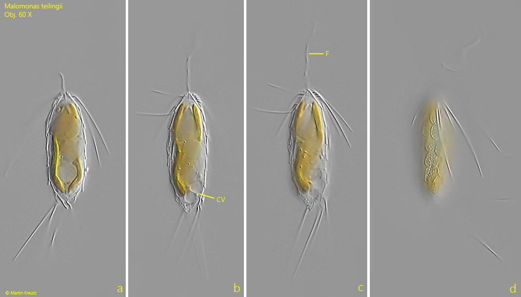
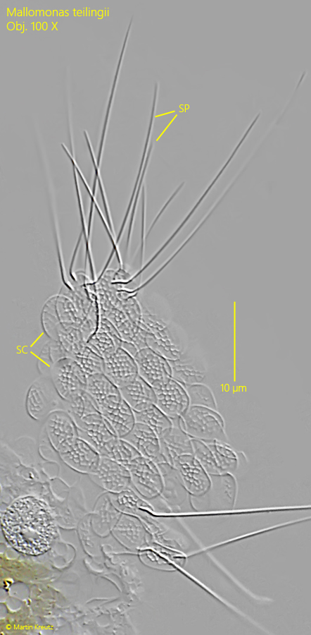
In February 2025, I found Mallomonas teilingii in the plankton of the Ziegelhof pond. The cells can be easily recognized by their slender shape and the tufts of long spines at both ends of the body. The scales are broadly elliptical and show a honeycomb pattern even under the light microscope (s. fig. 3). The spines are straight or slightly curved and smooth. When the cell contracts slightly, the spines can be folded toward the center of the body (s. figs. 1 c and 1 d).
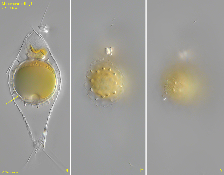
In addition to the living cells, I was also able to find numerous cysts, most of which were still enclosed in the empty shells of scales (s. fig. 4 a-c). The cysts had a diameter of 24–28 µm and were characteristically shaped by their covering of blunt, short spines.
Fig. 1 a-d:Mallomonas teilingii. L = 37 µm (without spines). A freely swimming specimen. CV = contractile vacuoles, F = flagellum. Obj. 60 X
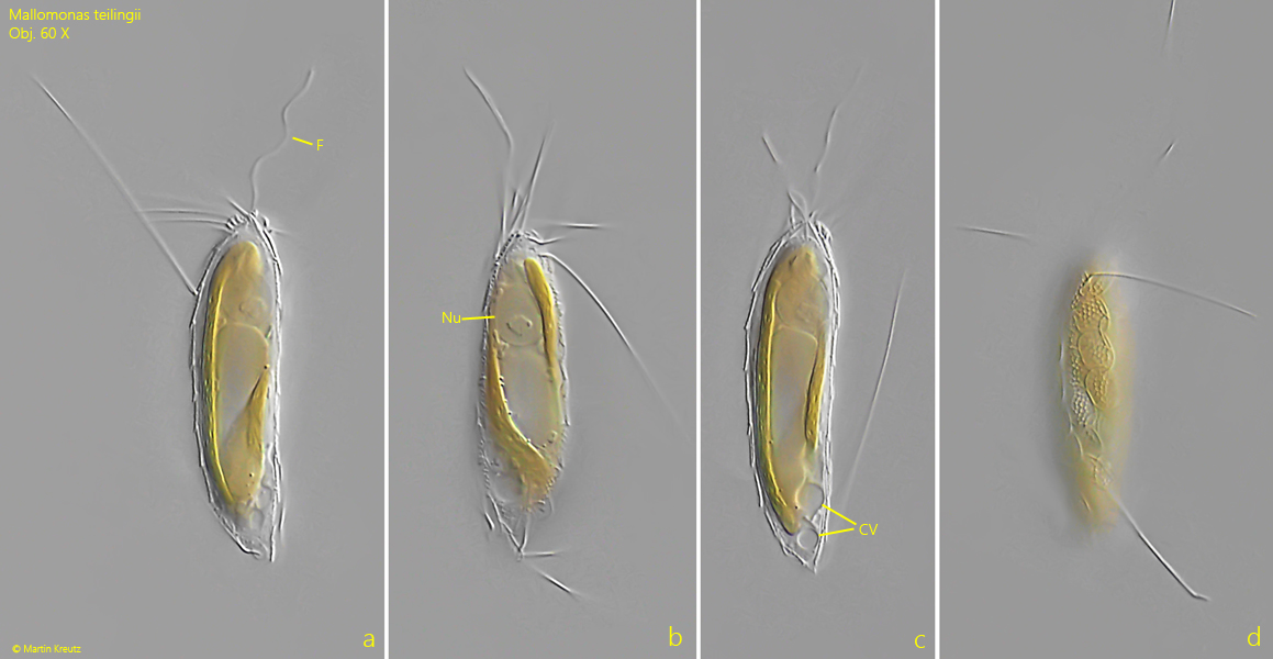
Fig. 2 a-d:Mallomonas teilingii. L = 48 µm (without spines). A second freely swimming specimen. CV = contractile vacuoles, F = flagellum, Nu = Nucleus. Obj. 60 X
Fig. 3:Mallomonas teilingii. The scales (SC) and spines (SP) of a strongly squashed specimen. Note the honeycomb-like pattern of the broadly elliptical scales. Obj. 100 X
Fig. 4 a-c:Mallomonas teilingii. The spherical cyst with a diameter of 27 µm in a empty shell of scales. The cyst is covered with blunt spines. Obj. 100 X