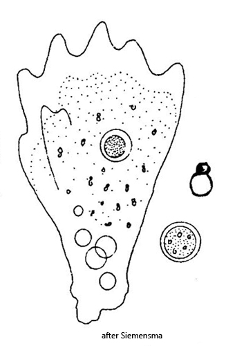
floating form about triangular, blunt anterior end
pseudopodia mamilliform or conical
length 35–125 µm
one contractile vacuole
one spherical nucleus (5.6–15 µm) with a large, central nucleolus
nucleolus sometimes with granular structure
small paired crystals in cytoplasm attached to spherical bodies
cuticula 0.18–0.2 µm thick layer
Mayorella penardi
Mayorella penardi is one of the most common naked amoeba species that I find in my samples. In fresh samples the specimens are often difficult to find, but Mayorella penardi likes to settle on the floating coverslip. There it is easy to observe.
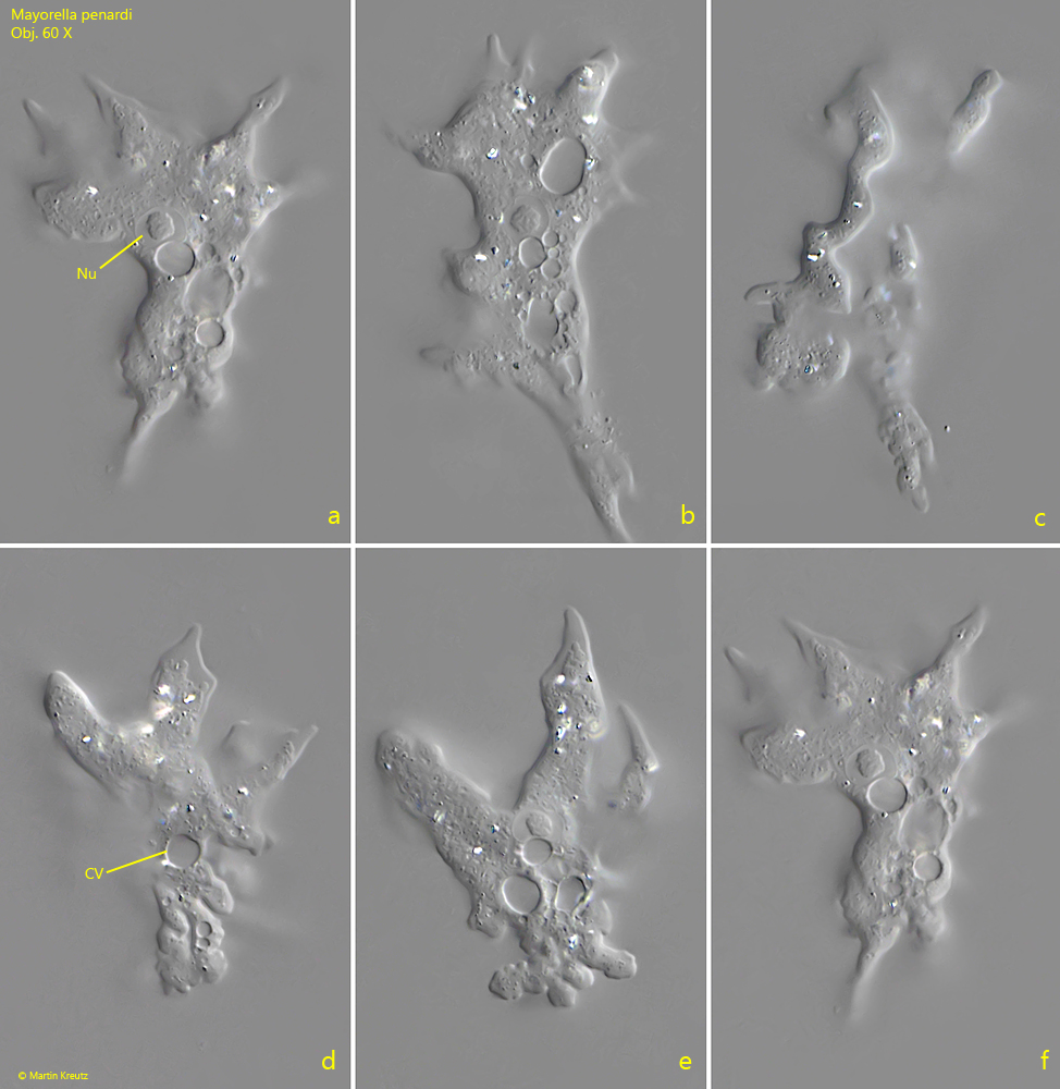
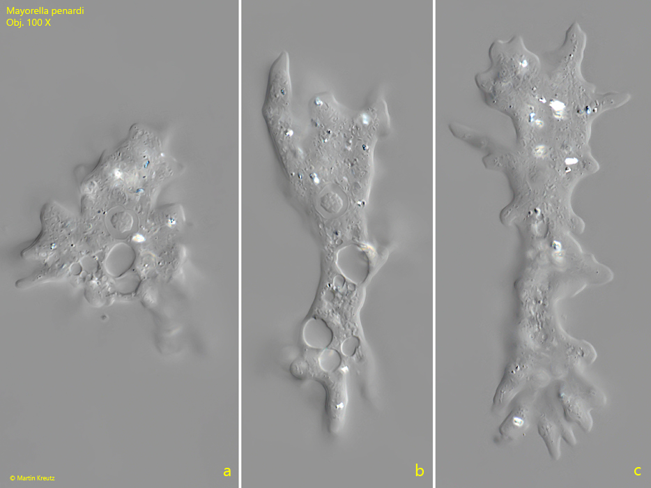
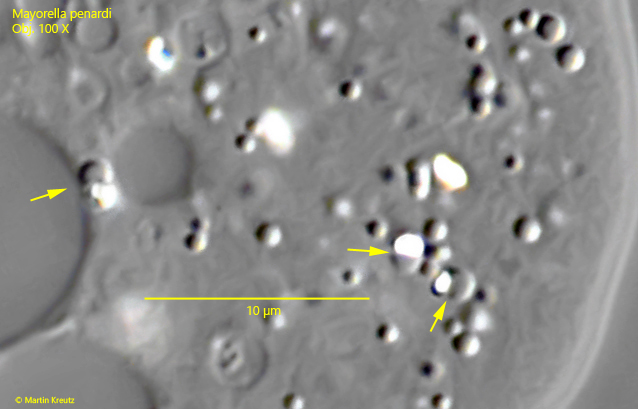
Most of the specimens are clearly under 100 µm in size. Only specimens that assume an elongated shape reach lengths of up to 120 µm. The cell nucleus is usually found near the middle of the body and has a distinct, round or knobby nucleolus, which can also be granular (s. fig. 1 a). The pseudopodia have the mamilliform shape typical of the genus Mayorella. That is, they taper more strongly toward the end and then almost take on a finger-like shape. In addition, the ends of the pseudopodia are always completely hyaline without inclusions. There is only one contractile vacuole, which is often surrounded by smaller auxiliary vacuoles. The colorless cytoplasm is mostly transparent with a few crystals that are coupled to oil-like droplets (s. fig. 4). This feature is also found in other Mayorella species, such as Mayorella augusta.
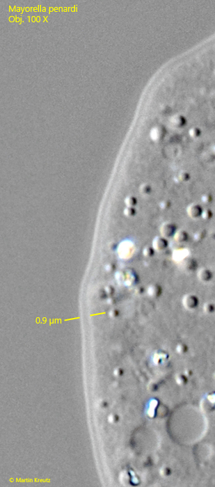
Another characteristic typical of the genus Mayorella is a thin cuticle that surrounds the entire cell body. This cuticle can be clearly seen at high magnification. Siemensma (s. link below) determined the thickness of the cuticle to be 0.18–0.2 µm. In my population it was noticeably thicker, at 0.8–1.0 µm (s. fig. 3).
Fig. 1 a-f:Mayorella penardi. L = 72–80 µm. A freely gliding specimen. Note the mamilliformed pseudopodia. CV = contractile vacuole, Nu = nucleus. Obj. 60 X.
Fig. 2 a-c:Mayorella penardi. L = 60–116 µm. A contracted (a) and elongated (b, c) second specimen. Obj. 100 X.
Fig. 3:Mayorella penardi. The cell is covered by a thin cuticula. In this specimen the layer is 0.9 µm thick. Obj. 100 X.
Fig. 4:Mayorella penardi. In the cytoplasm highly refractive crystals are present, paired with small, oily droplets (arrows). Obj. 100 X.
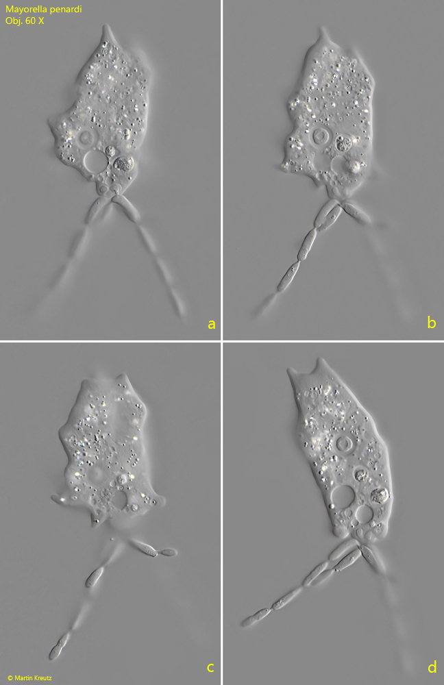
In very old samples in particular, which have been left to stand for several weeks, I more frequently find specimens of Mayorella penardi that are infected by an exoparasitic fungus. This is expressed in the fact that the infected specimens trail a segmented thread behind them (s. fig. 1 a-d). Usually there are two to three of these threads.
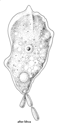
The fungus is Amoebophilus simplex (s. figs. 6 and 7), which is host-specific, meaning it infects exclusively Mayorella penardi. The segmented threads are the conidia of the fungus. Mature cells of the conidia break off at the ends of the threads and are then phagocytosed by further specimens of Mayorella penardi that are not yet infected. In this way Amoebophilus simplex reproduces.
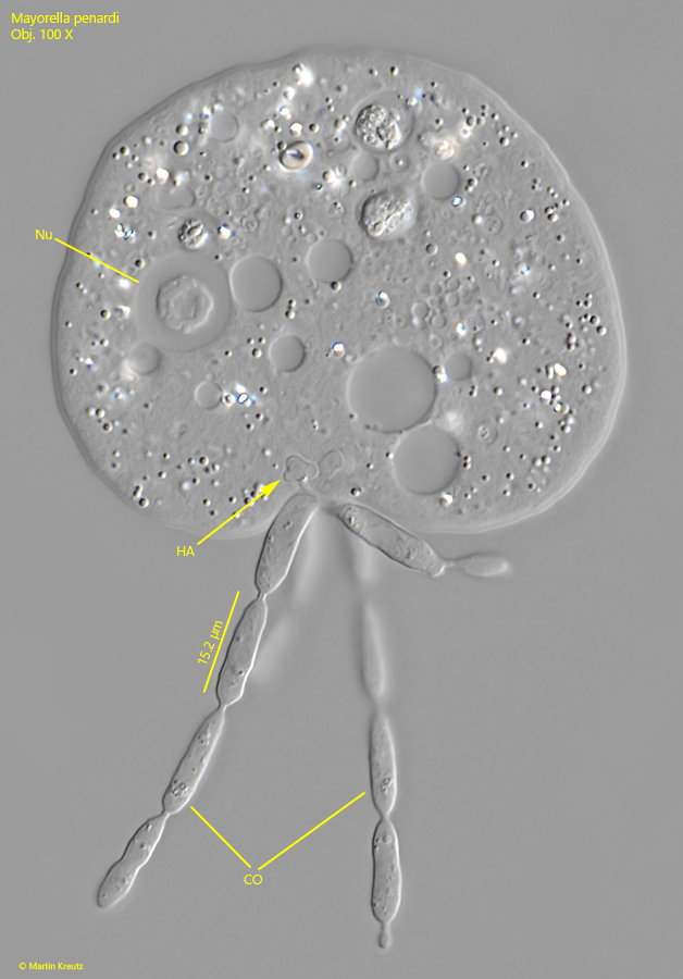
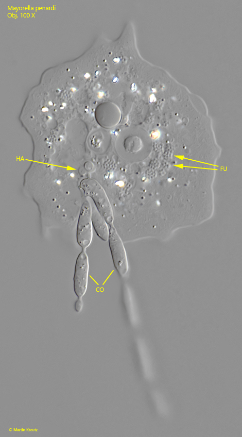
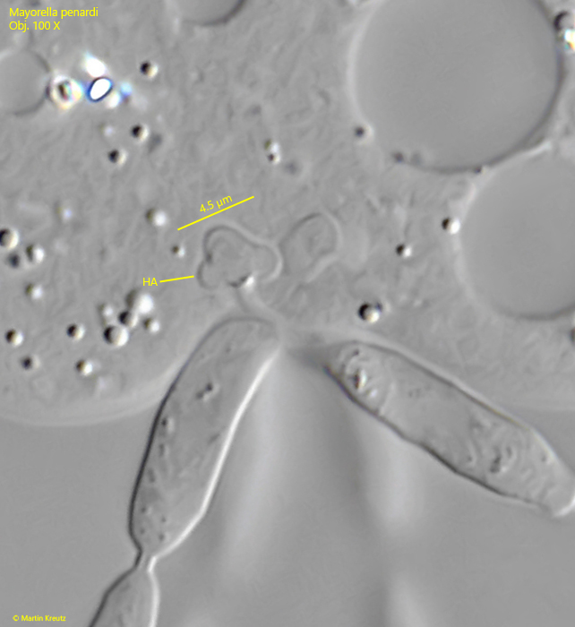
The conidia cannot be stripped off by the amoeba, which is always astonishing to observe. In addition, they are always located at the posterior end of the amoeba, opposite to the direction of movement. The anchoring of the conidia takes place via so‑called haustoria. These are specialized cells which, in Amoebophilus simplex, have a cloverleaf or tuberous shape (s. figs. 8, 9 and 10). The haustorium absorbs nutrients from the cytoplasm of the host cell and cannot simply be expelled by the host cell, which results in the anchoring. How the haustorium manages not to be expelled by the host cell through an invagination of the cell membrane is unknown.
Mrva (2008) and Siemensma (s. link below) report that specimens of Mayorella penardi infected with Amoebophilus simplex are found only extremely rarely. In my experience, the likelihood of finding infected specimens increases with the age of the samples. Apparently, the fungus can reproduce much more effectively in this limited habitat. Incidentally, this also applies to other cases of parasitism, regardless of whether they involve euglenids, ciliates, or rotifers.
Fig. 5 a-d:Mayorella penardi. L = 66–72 µm. A specimen infested by an exoparasitic fungus. The thread-like conidia of the fungus arise from the uroid. Obj. 60 X.
Fig. 6:Mayorella penardi. Drawing of a specimen infested by the exoparasitic fungus Amoebophilus simplex.
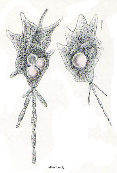
Fig. 7: These drawings by Leidy from the year 1879 very likely show Mayorella penardi, infected by Amoebophilus simplex. The conidia were misinterpreted by Leidy as part of the uroid.
Fig. 8:Mayorella penardi. The squashed specimen as shown in fig. 5 a-d with the segmented conidia (CO) of Amoebophilus simplex. The segments of the conidia are about 15 µm long. The conidia are anchored with a special shaped haustorium (HA) in the host cell. Obj. 100 X.
Fig. 9:Mayorella penardi. A second squashed specimen infested by Amoebophilus simplex. In the cytoplasm further fungal cells (FU) are visible, likely a stage of the life cycle of Amoebophilus simplex. CO = conidia, HA = haustorium. Obj. 100 X.
Fig. 10:Mayorella penardi. A cloverleaf-shaped haustorium (HA) of Amoebophilus simplex in detail. This organell is specialized to absorb nutrients from the cytoplasm of the host cell. The haustorium cannot simply be expelled from the host cell through the cytoplasm membrane, which results in anchoring. Obj. 100 X.