Mesostigma grande Korshikov, 1938
Most likely ID: n.a.
Synonym: n.a.
Sampling location: Pond of the waste disposal company Constance
Phylogenetic tree: Mesostigma grande
Diagnosis:
- cells obvoid, compressed, saddle-shaped
- length about 27 µm, width 18–20 µm, thickness 8 µm
- cell covered by gelatinous sheath with rod-shaped scales
- two short flagella arise on convex side from shallow depression
- 3–7 contractile vacuoles near depression
- chloroplast cup-shaped
- one large eyespot on concave side near equator
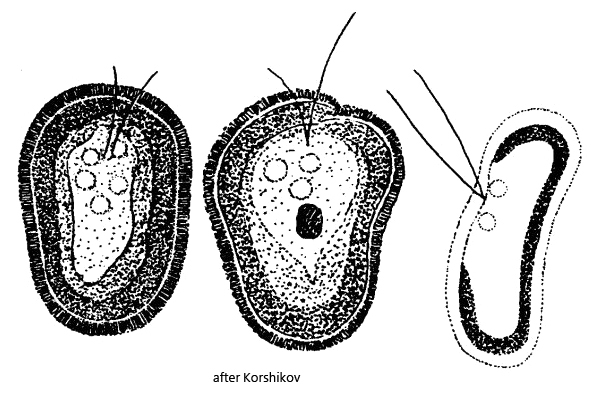
In July 2026, I found several specimens of Mesostigma grande in a plankton sample from the pond of the waste disposal company Constance. The pond is highly eutrophied.
There are only a few records and observations of Mesostigma grande. The descriptions by Korshikov, who found the species in 1938 in a canal in southern Russia, were adopted by later authors such as Huber-Pestalozzi (1961).
Mesostigma grande is easy to identify. The cell is flattened and saddle-shaped with two strongly arched edges. There is thus a concave and a convex side. In addition, the cell is covered by a distinct layer of scales, which were described by Korshikov as short rods standing perpendicular to the cell surface. The eyespot is located almost in the middle of the cell. It is brick-red, large, and irregularly shaped (s. fig.s 1 f and 2 f). The two flagella are of equal length and not longer than the body (s. fig. 1 a). Where the flagella arise, there are also several contractile vacuoles with varying rates of activity (s. fig. 2 e).
In my population I was able to observe some deviations from Korshikov’s description. The shape of the cells was quite different and not always obvoid. They were often somewhat asymmetrical and rather rectangular. In addition, their size was quite variable, between 20–30 µm. According to my observations, the two flagella originate from the concave side and not the convex one. In contrast, the eyespot is located on the convex side and not on the concave, thus exactly the other way round from how Korshikov described it. Both the eyespot and the nucleus were arranged near the middle of the body in all specimens. The flagella originated in the anterior third. I was not able to discern the depression from which they are supposed to arise.
The scales appear to be shaped differently than described by Korshikov. They are not simply short rods, but seem to possess a hemispherical base that sits on the cell wall, and on the side facing away they bear a short spine. The short spines can be clearly seen (s. fig. 3). That the base of the scales must be round is recognized when one focuses on them. Then it looks as if small discs are arranged closely adjacent to one another (s. fig. 3). The exact shape of the scales can probably only be determined by electron microscopy.
Mesostigma grande can be reliably distinguished from the similar species Mesostigma viride by various characteristics. At 15–18 µm in length, Mesostigma viride is about one third smaller. In addition, this species does not possess a layer of scales, and the chloroplast often contains 1–2 pyrenoids.
Huber-Pestalozzi still regarded Mesostigma grande as a volvocale alga, which seems plausible in view of the eyespot, contractile vacuole, and flagella. On the basis of genetic studies by Bhattacharya et al. (1998), the genus Mesostigma was considered the most primitive representative of the Streptophyta. This classification was questioned again in 2001 by Karol et al. As long as no decision has been made, I will leave Mesostigma grande among the Volvocales.
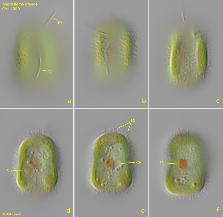
Fig. 1 a-f: Mesostigma grande. L = 21 µm. Different focal planes of a freely swimming specimen from the concave side. Note the layer of scales (LS) covering the cell. CV = contractile vacuole, ES = eyespot, F1/F2 = flagella, Nu = nucleus. Obj. 100 X.
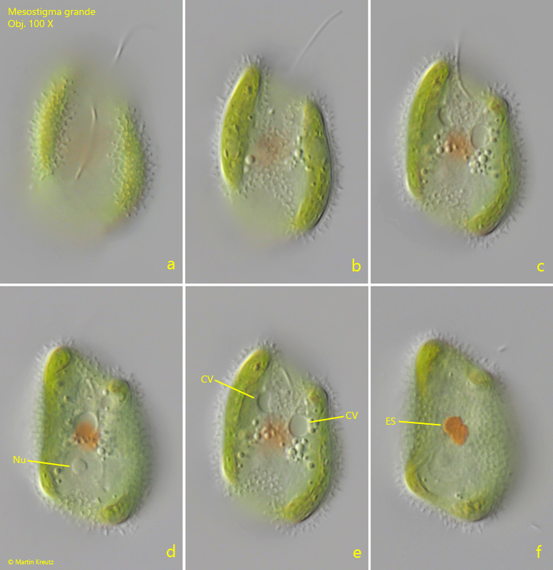
Fig. 2 a-f: Mesostigma grande. L = 29 µm. Different focal planes of a second, larger specimen. This specimen ist shaped asymmetric and has several contractile vacuoles (CV). ES = eyespot, Nu = nucleus. Obj. 100 X.
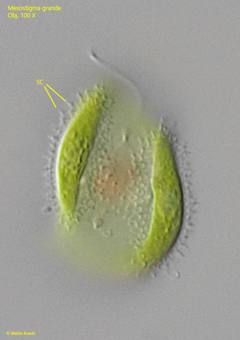
Fig. 3: Mesostigma grande. L = 29 µm. The scales (SC) in detail. It seems as if they have a hemispherical base with a small spike on it. Depending on the focal plane, the circular bases of the hemispheres can also be seen. The total length of the scales is 2.2 µm. Obj. 100 X.