Mesostigma viride

Lauterborn, 1894

Most likely ID: n.a.


Synonym: n.a.


Sampling location: Pond of the waste disposal company Constance 


Phylogenetic tree: Mesostigma viride


Diagnosis: 

  • cells obvoid or rhomboid, compressed, saddle-shaped
  • length 15–18 µm, width 11–13 µm, thickness 5–7 µm
  • cell covered by delicate, beaker-shaped scales
  • two short flagella arise on convex side from a pit
  • 3–7 contractile vacuoles near origin of flagella
  • chloroplast cup-shaped, yellowish-green
  • one large eyespot on concave side near equator
Mesostigma-viride
Mesostigma viride

In July 2026, I found several specimens of Mesostigma viride in plankton samples from the pond of the waste disposal company Constance. The pond is heavily eutrophicated.


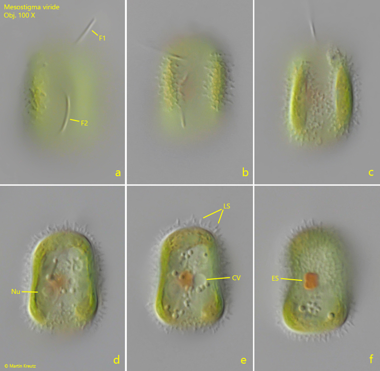
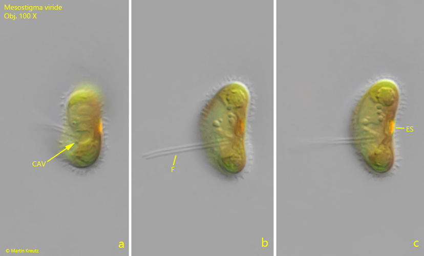
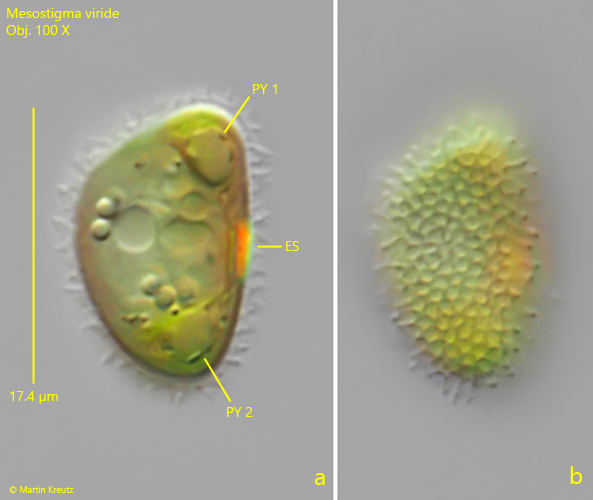
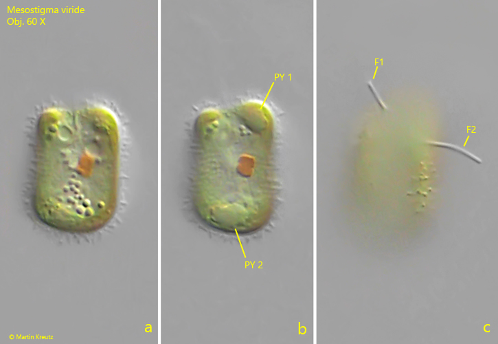
Mesostigma viride has an unusual body structure. The cells are not flattened laterally, but from the cell poles. That is, the anterior and posterior ends are brought together. As a result, the flagella and the eyespot are now more or less in the middle of the flattened disc. In addition, this disc is curved in the shape of a saddle, with two upturned edges. Thus, the saddle shape has a concave and a convex side. The two flagella of Mesostigma viride arise from a depression on the convex side, while the large brick-red eyespot lies approximately in the middle of the concave side (s. fig. 4 a-c). The chloroplast is yellow-green in color. Two pyrenoids are located at opposite ends of the cell (s. figs. 1 b and 5 a).


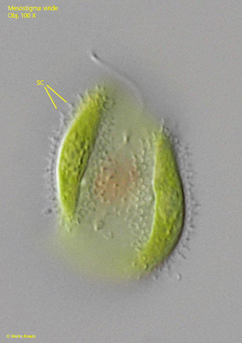
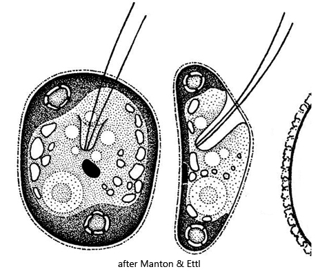
The periplast of Mesostigma viride is covered by a clearly visible layer of scales. The form of these scales was examined by electron microscopy by Manton & Ettl (1965). They are essentially cup-shaped and sit on a base of plate-like scales. Under the light microscope, the scales appear to consist of short spines (s. figs. 5 b and 6). However, these are the adjoining walls of the “cups.” In apical view, the scales appear approximately circular and lie close together.


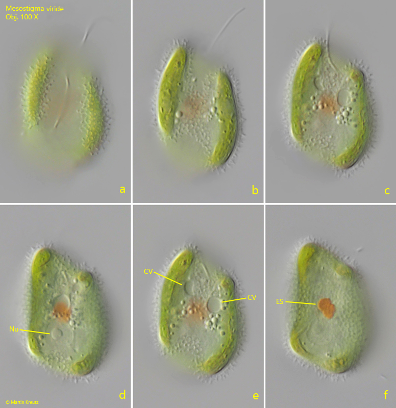
In my population, I was able to find cells 18–30 µm in length, which is considerably larger than the range of 15–18 µm given by Huber-Pestalozzi (1961). However, culture experiments by Manton & Ettl (1965) have shown that cell dimensions can be variable. The cells kept in culture were only 7.7–12.5 µm long. The size of the cells in my population may also be attributable to the highly eutrophic habitat.

The similar species Mesostigma grande can be distinguished from Mesostigma viride by the two pyrenoids, which are absent in Mesostigma grande. In addition, according to the description by Korshikov (1938), the scale layer of Mesostigma grande is said to consist of thin rods that stand perpendicular to the cell surface. In my opinion, the size of Mesostigma grande, approximately 27 µm, can no longer serve as a distinguishing characteristic from Mesostigma viride, since this is evidently variable and influenced by environmental conditions.


Huber-Pestalozzi (1961) still regarded Mesostigma viride as a volvocalean alga, which seems plausible given its eyespot, contractile vacuole, and flagella. Based on genetic studies by Bhattacharya et al. (1998), the genus Mesostigma was considered the most basal representative of the Streptophyta. This classification was questioned again in 2001 by Karol et al. Until a decision has been reached, I will leave Mesostigma viride in the Volvocales.
 
More images and information on Mesostigma viride: Dr. Bernd Laber -Mikro-Forum-Mesostigma viride-Eine ungewöhnliche Alge (registration required)
Mesostigma-viride

Fig. 1 a-c: Mesostigma viride. L = 20 µm. Different focal planes of a freely swimming specimen from the concave side. Note the two pyrenoids (Py 1, PY 2) and two flagella (F1, F2). Obj. 60 X.

Mesostigma-viride

Fig. 2 a-f: Mesostigma viride. L = 21 µm. Different focal planes of second freely swimming specimen from the concave side. Note the layer of scales (LS) covering the cell. CV = contractile vacuole, ES = eyespot, F1/F2 = flagella, Nu = nucleus. Obj. 100 X.

Mesostigma-viride

Fig. 3 a-f: Mesostigma viride. L = 29 µm. Different focal planes of a third, larger specimen. This specimen ist shaped asymmetric and has several contractile vacuoles (CV). ES = eyespot, Nu = nucleus. Obj. 100 X.

Mesostigma-viride

Fig. 4 a-c: Mesostigma viride. L = 18 µm. In lateral view the cavity (CAV) on the convex side is visible, where the two flagella (F) arise. The eyespot (ES) is located on the concave side. Obj. 100 X.

Mesostigma-viride

Fig. 5 a-b: Mesostigma viride. L = 18 µm. The same specimen as shown in fig. 4 a-c with focal plane on the pyrenoids (PY 1, PY 2) and on the scales covering the periplast (b). Obj. 100 X.

Mesostigma-viride

Fig. 6: Mesostigma viride. L = 29 µm. The scales (SC) in detail. Depending on the focal plane, the circular bases of the scales can also be seen. The thickness of the layer of scales is 2.2 µm. Obj. 100 X.