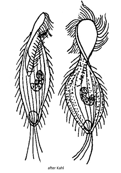
macronucleus kidney-shaped or ellipsoidal with adjacent micronucleus
contractile vacuole large, terminal
posterior end with caudal cilia
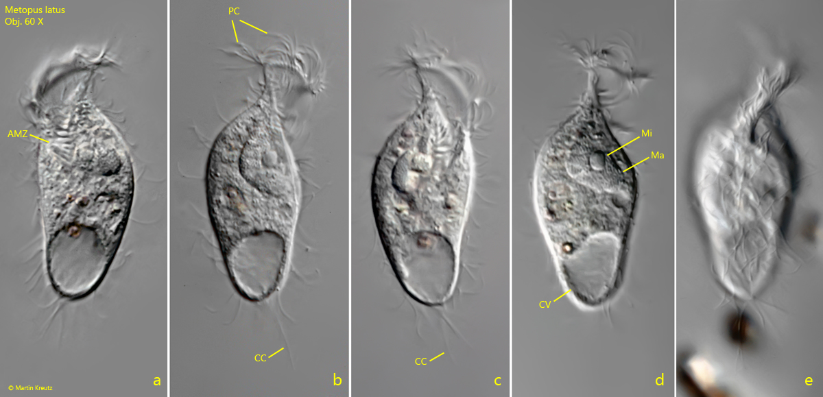
Metopus latus
So far I could find only one specimen of Metopus latus in October 2006 in the Simmelried. The photos shown below were still taken on slide film with a 60 X lens at high layer thickness.
Metopus latus can be easily recognized by the flattened and twisted anterior dome, on the outer edge of which runs the perizonal stripe with long cilia (s. fig. 1 b). The adoral zone is only short and has few membranelles (s. fig. 1 a). Kahl gives a length of about 85 µm. My specimen was a bit stouter and only 68 µm long, but this is still within the usual variability. As described and drawn by Kahl (s. above), the macronucleus is kidney-shaped and encloses a spherical micronucleus (s. figs. 1 b and 1 d). The caudal cilia are long but widely spaced.
Fig. 1 a-e:Metopus latus. L = 68 µm. A freely swimming specimen from ventral (a, c) and from right (b, d, e). Note the flattened and twisted apical dome with the long perizonal cilia on the edge. AMZ = adoral zone of membranelles, CC = caudal cilia, CV = contractile vacuole, Ma = macronucleus, Mi = micronucleus. Obj. 60 X.