I find Micrasterias crux-melitensis more frequently at the sampling sites in my area. The specimens are either on the top layer of mud or as growth on aquatic plants.
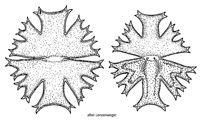
Micrasterias crux-melitensis can be easily recognized by the two apical lobes, which surpass the lateral lobes and are separated from them by wide, V-shaped notches. The sinus, which separates both semi-cells, is also widely open.
Fig. 1 a-b:Micrasterias crux-melitensis. L = 129 µm. Two focal planes of a slightly squashed specimen. Obj. 60 X.