semicells 3-lobed, with the 2 interlobular incisions
lobes projecting horizontally
walls minutely punctate
Micrasterias pinnatifida
I found Micrasterias pinnatifida in 1996 in the Determoor (Austria) and a second time in 2025 in the Schwemm Moor (Austria). In the samples from the Schwemm Moor, Micrasterias pinnatifida occurred quite frequently. The specimens shown below come from the Schwemm Moor.
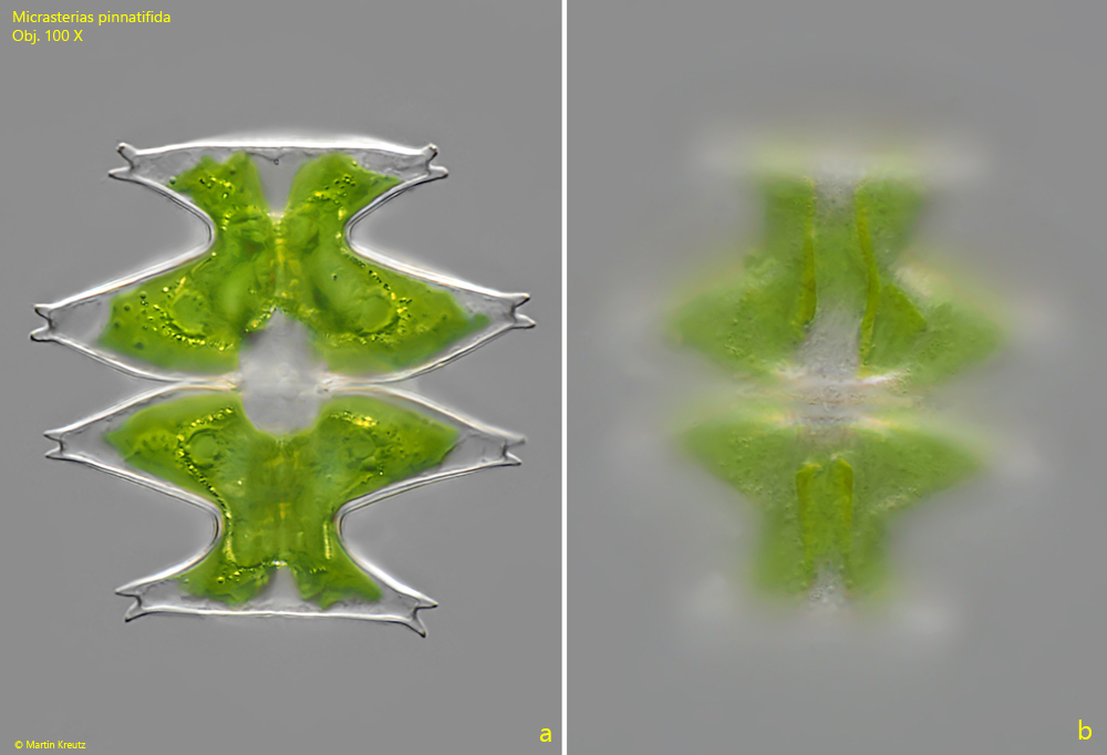
Micrasteris pinnatifida is notable for its small size and comparatively simple segmentation of the lobes. Each semi-cell is three-lobed. The lobes each bear a pair of short spines at their ends. The chloroplasts contains “several” pyrenoids each (Förster, 1982). In my population there were mostly 4 pyrenoids per semi-cell.
Fig. 1 a-b:Micrasterias pinnatifida. L = 58 µm. A slightly squashed specimen in DIC. Obj. 100 X.
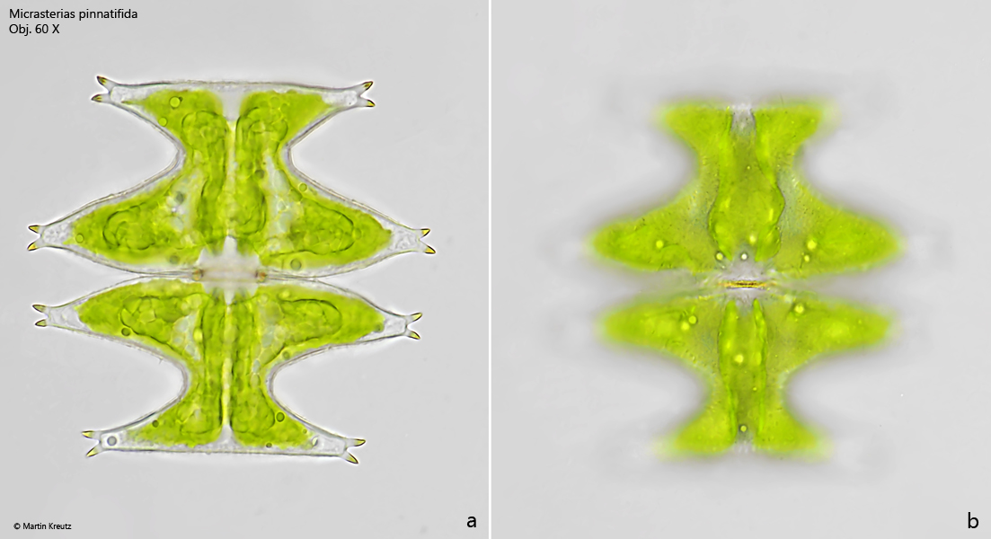
Fig. 2 a-b:Micrasterias pinnatifida. L = 76 µm. A second specimen in brightfield illumination. Obj. 60 X.