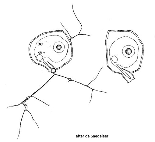
shell retort-shaped, outline circular, angular or asymmetrical
length of shell 12–21 µm
neck with a septum
neck follows more or less the shape of the shell, no collar perpendicular to shell outline
protoplast fills the shell only partially
nucleus central with a spherical nucleolus
contractile vacuole near neck
granuloreticulopodia very thin and anastomosing, arising from a peduncle
Microgromia longisaepimen
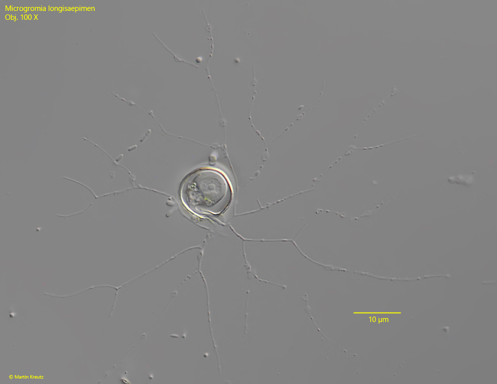
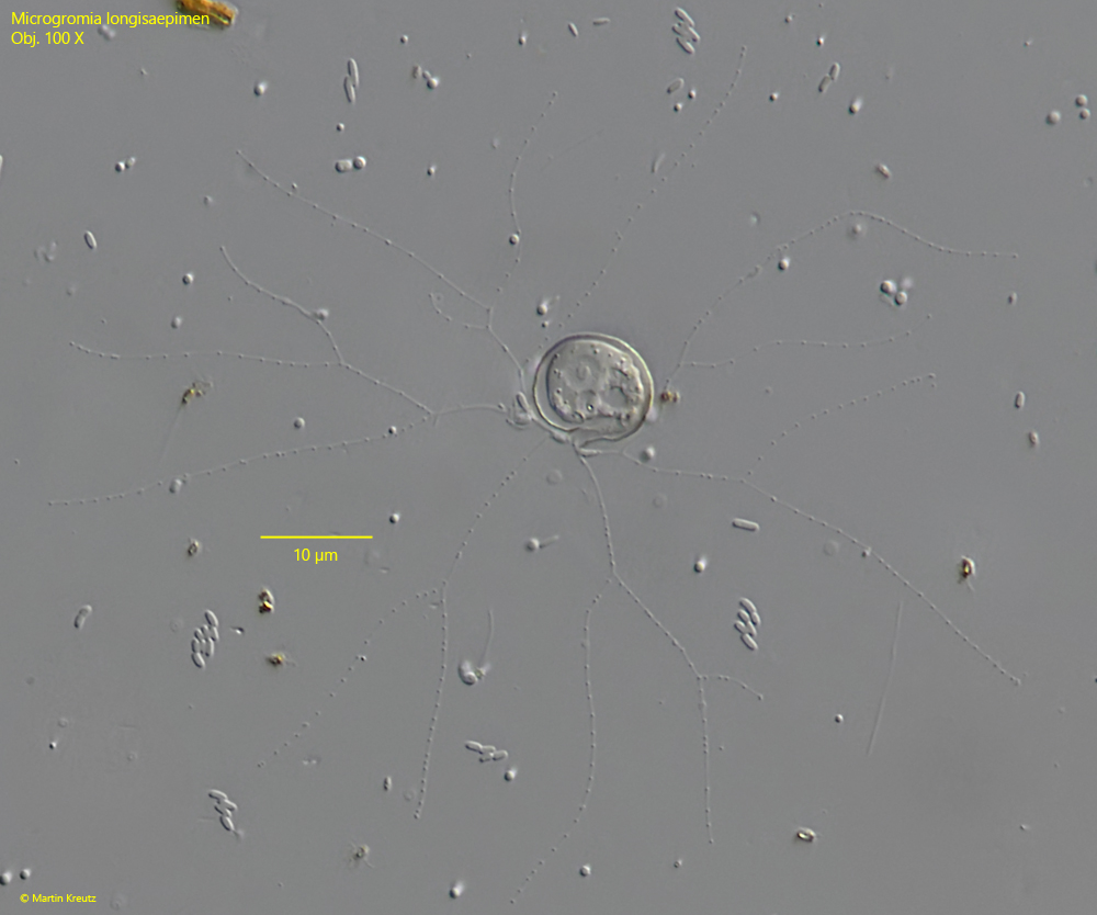
I find Microgromia longisaepimen exclusively in the Simmelried. There I find this testate amoeba reliably and often. However, in most cases it is cannot observed directly in the samples but can easily be isolated from the detritus by floating coverslips and then observed very well. During reproduction Microgromia longisaepimen forms flagellated swarmers, which settle on the coverslips after only a few days. On the coverslips the fine granuloreticulopodia spread widely to collect bacteria, small algae or even small diatoms (s. figs. 1, 2 and 4).
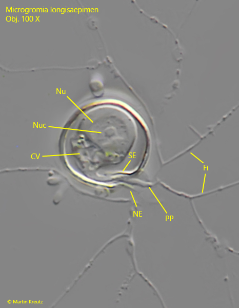
Microgromia longisaepimen can be distinguished from the other Microgromia species by the shape of the neck. It follows the shape of the shell as in a snail shell and is separated from the shell lumen by a clearly visible septum (s. fig. 2).
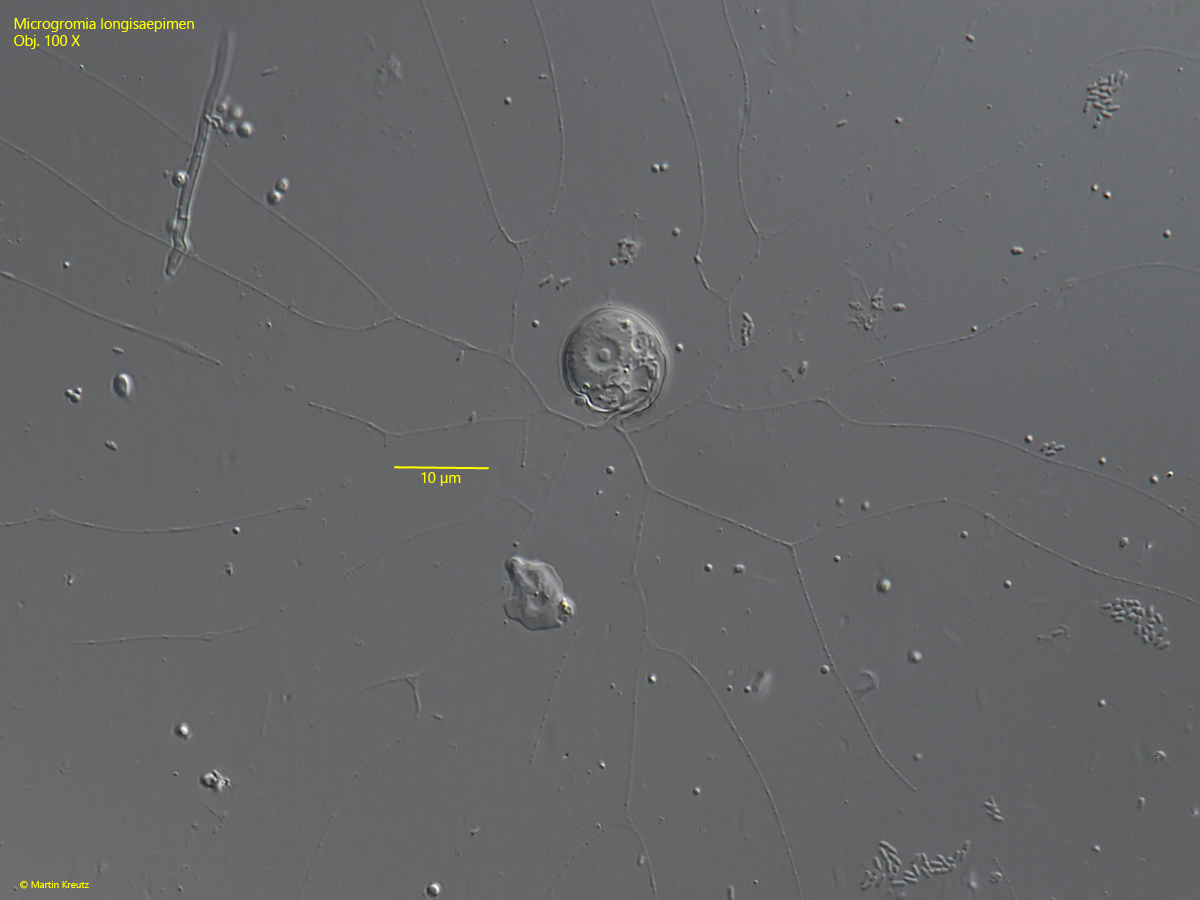
Fig. 1:Microgromia longisaepimen. L = 12 µm. A fully expanded specimen. Note the widely branched granuloreticulopodia. Obj. 100 X.
Fig. 2:Microgromia longisaepimen. L = 12 µm. The same specimen as shown in fig. 1 in detail. CV = contractile vacuole, Fi = filopodia with granula (granuloreticulopodia), Nu = nucleus, Nuc = nucleolus, NE = neck, PP = cytoplasm peduncle, SE = septum. Obj. 100 X.
Fig. 3:Microgromia longisaepimen. L = 12 µm. A second fully expanded specimen. Obj. 100 X.
Fig. 4:Microgromia longisaepimen. L = 13 µm. A third fully expanded specimen. Obj. 100 X.
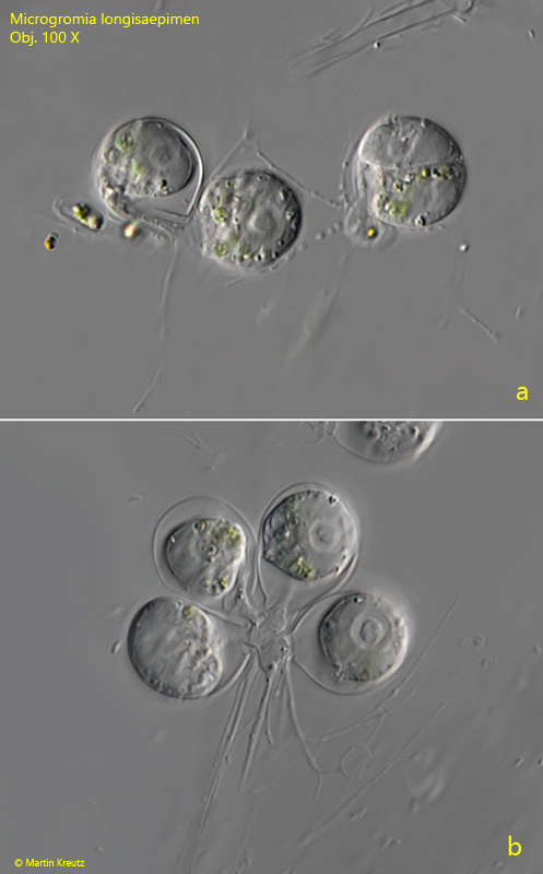
In some samples I could observe feeding communities of Microgromia longisaepimen (s. fig. 5a-b). The food consisted of small algae or cyanobacteria. The plasma peduncles of the individual specimens fuse and then form a common network of granuloreticulopodia with which the food particles are collected.
Fig. 5 a-b:Microgromia longisaepimen. Two feeding communities of three (a) and four specimens (b). In a feeding community the cytoplasmic peduncles fuse to collect food and to share it (b). Obj. 100 X.
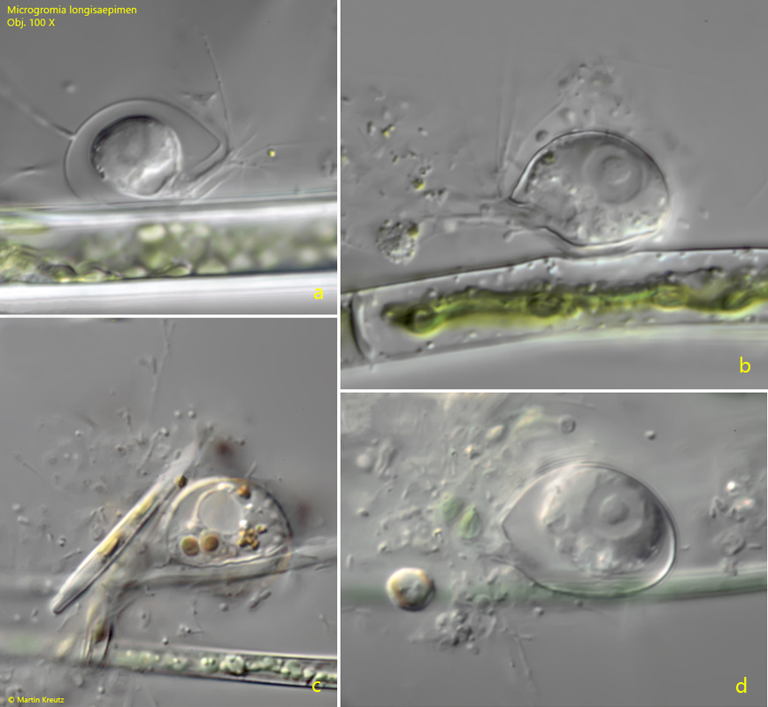
Microgromia longisaepimen can attach itself to algal filaments or other solid materials. This property can be used to grow specimens on floating coverslips. In natural environments, algal threads are often colonized. In this case, the housing encloses the algal filament, similar to a cramp.
Fig. 6 a-d:Microgromia longisaepimen. Several specimens attached to algae filaments. To hold on to an algal filament, the shell partially grows around it, similar to a cramp (c). Obj. 100 X.