ventral margin almost straight with indentation near posterior end
left side with three distinct ribs
spherical macronucleus in center of cell
one spherical micronucleus
contractile vacuole in posterior third
three ciliary rows on right side parallel to the convex dorsal margin
oral apparatus at posterior end near ventral margin
Microthorax costatus
So far I have only found Microthorax costatus in the Simmelried. However, the records are very sparse. On average, I find a few specimens every 4 years. Because the mall size of the species, it is possible that I have overlooked finds.
Microthorax costatus is usually found on gelatinous colonies of bacteria, rhodobacteria or cyanobacteria, which are obviously being grazed. In the literature I have only found the very brief description and drawings by Kahl (1926), who also found Microthorax costatus only occasionally and described it first. Obviously there are no further records of this species after that.
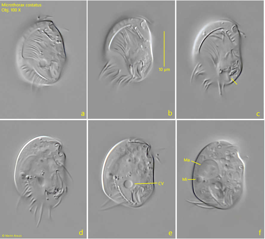
With a length of 16-22 µm, my specimens were somewhat smaller than described by Kahl (about 25 µm). Very characteristic, however, are the 3 ribs on the left side, which end nose-shaped towards the posterior end (s. figs. 3 a-c and 4 a-c). In addition, the end of the middle rib is slightly bent ventrally. The right side has the typical structure of the genus Microthorax. Three longitudinal rows of cilia run parallel to the convex posterior margin. The two dorsal rows are interrupted in the middle. The third row is only rudimentary and very short. It is difficult to recognize. The cilia of these rows are conspicuously long (s. figs. 1 b and 1 d). The macronucleus is round and quite large. I was able to recognize a spherical micronucleus adjacent to the macronucleus (s. figs. 1 f and 2 b). The contractile vacuole is located in the posterior third near the oral apparatus (s. fig. 1 e).
Fig. 1 a-f:Microthorax costatus. L = 19.6 µm. Different focal planes of a freely swimming specimen from right. CV = contractile vacuole, Ma = macronucleus, Mi = micronucleus. Obj. 100 X.
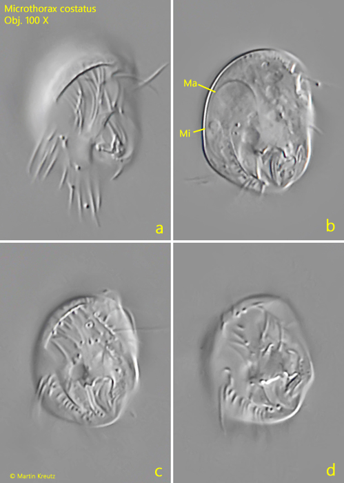
Fig. 2 a-d:Microthorax costatus. L = 18 µm. Different focal planes of a second specimen from right. Ma = macronucleus, Mi = micronucleus. Obj. 100 X.
Fig. 3 a-c:Microthorax costatus. L = 19 µm. Different focal planes of the left side with the three distinct ribs (arrows). Ma = macronucleus. Obj. 100 X.
Fig. 4 a-c:Microthorax costatus. L = 22 µm. A second specimen from the left side. Obj. 100 X.