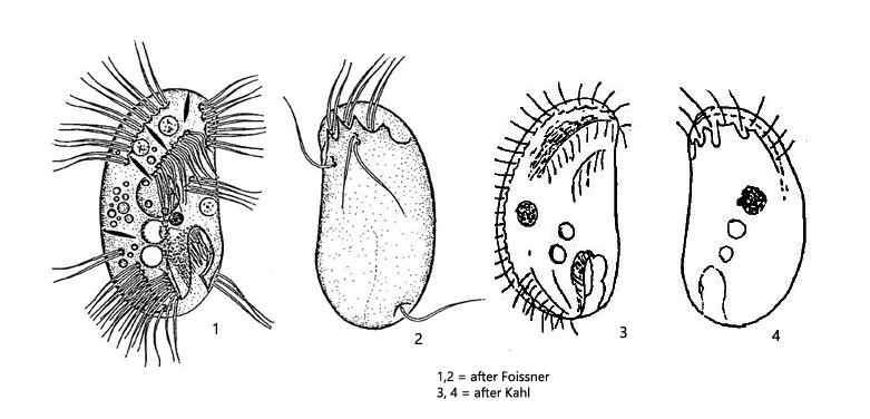
Microthorax pusillus is a very common ciliate in my sites Ulmisried and Simmelried. There I find this ciliate in the mud or between decomposing plant masses. In my populations some specimens with 40 µm length were about 10 % larger than given by Kahl and Foissner (s. fig. 3 a-b). Although the ciliate is quite small, it is easily recognized by the bean-shaped body and the mouth opening located at the posterior end. On the right side of the body there are 3 rows of cilia, which are interrupted in the middle of the body (s. fig. 4a). Each cilium in these rows originates in a separate depression in the pellicle, creating a typical chain pattern.
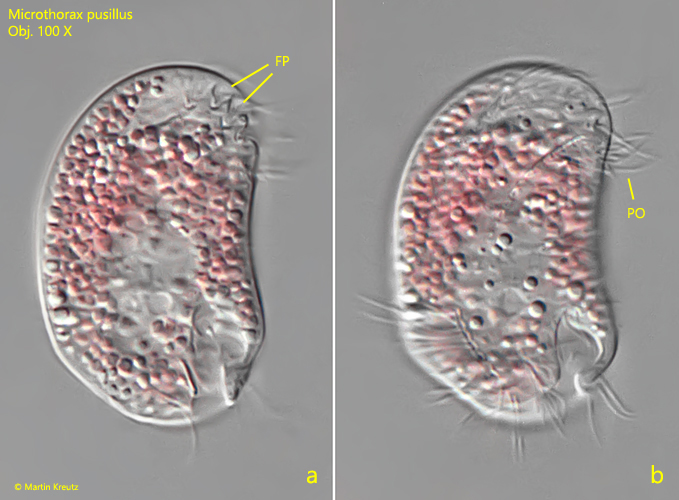
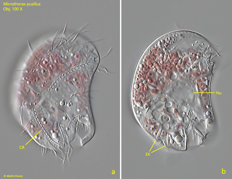
Microthorax pusillus feeds on bacteria but also on purple bacteria. Since the Simmelried is rich in purple bacteria, I have found several times specimens here, which obviously fed preferentially on purple bacteria (s. figs. 3 a-b and 4 a-b). Thus the specimens also appeared intensely pink at low magnification. At high magnifications, partially decomposed purple bacteria were then visible in the cytoplasm due to the ongoing digestion process.
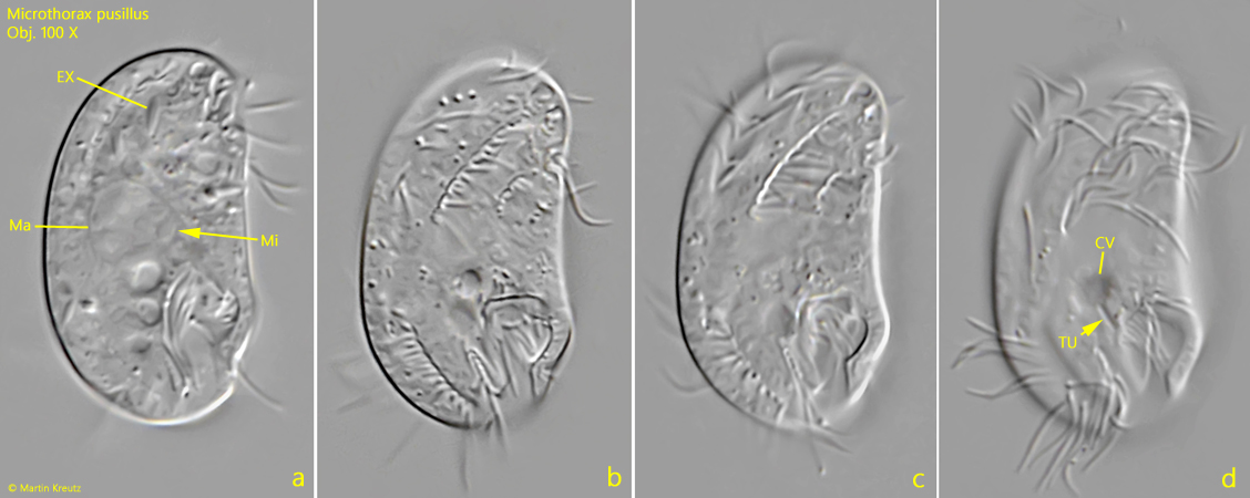
Fig. 1 a-d:Microthorax pusillus. L = 28 µm. Different focal planes of a freely swimming specimen from right. Note the short tube (TU) between the contractile vacuole (CV) and the oral apparatus. EX = extrusomes, Ma = macronucleus, Mi = micronucleus. Obj. 100 X.
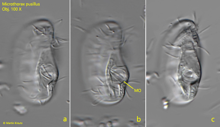
Fig. 2 a-c:Microthorax pusillus. L = 25 µm. Three focal planes of the mouth opening (MO). Obj. 100 X.
Fig. 3 a-b:Microthorax pusillus. L = 40 µm. Two focal planes of the right side of a specimen that fed on purple bacteria found in March 2003. FP = furrows of praeoral cilia rows, PO = praeoral cilia. Obj. 100 X.
Fig. 4 a-b:Microthorax pusillus. L = 38 µm. Two focal planes of the right side of a second specimen that fed on purple bacteria found in June 2022. CR = interrupted cilia rows of right side, EX = extrusomes, Nu = nucleus. Obj. 100 X.