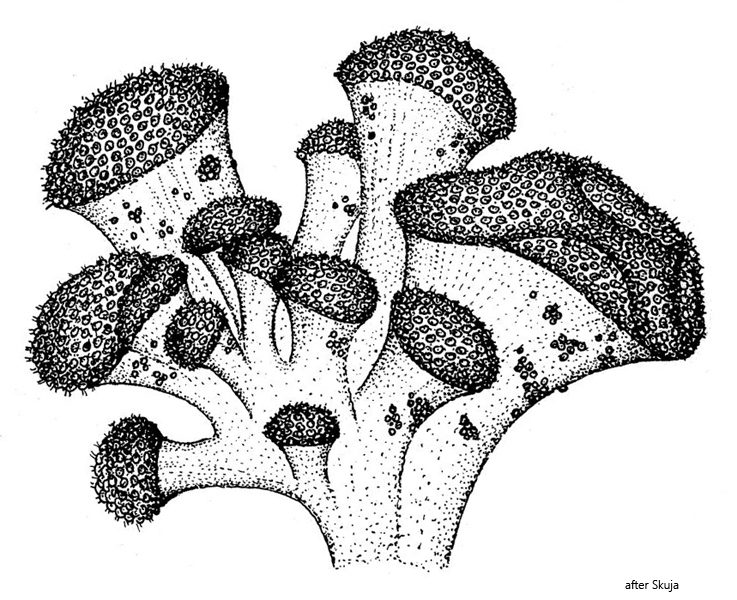
I find colonies of Mycochrysis oligothiophila very rarely in the Simmelried. Most of the time the colonies are located among submerged and decomposing aquatic plants. All colonies that I have found so far were unbranched but possessed the typical shape of a mushroom cap. This could be because Mycochrysis oligothiophila is adapted to habitats with a high concentration of hydrogen sulfide, which is only conditionally the case in the Simmelried. In my other locations I have never found this species.
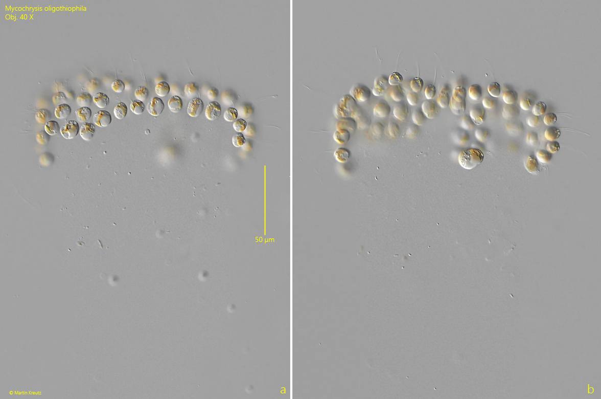
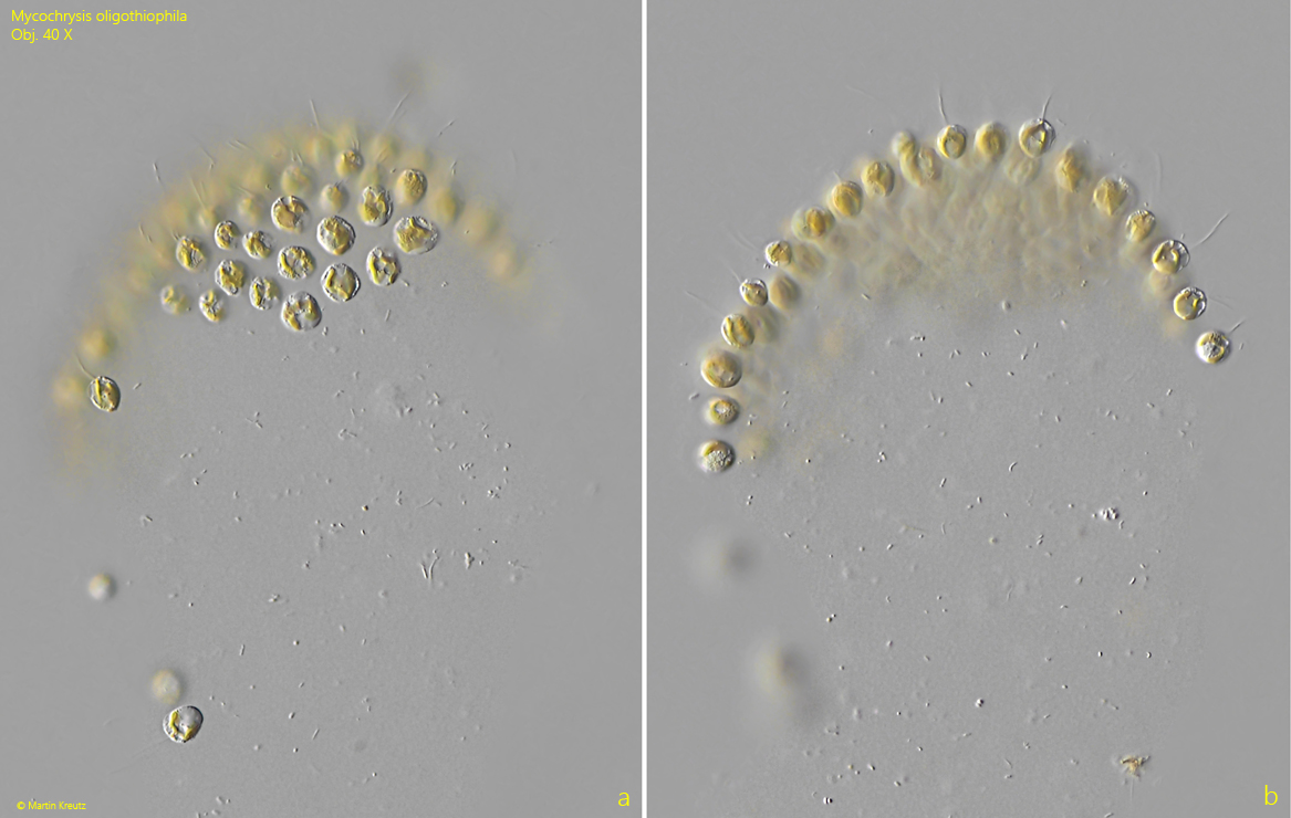
The broad stalks of the colonies are gelatinous and appear slightly granular in DIC and are difficult to bring into contrast (s. fig. 1 a-b). The shape of the stalks is best recognized by adhering bacteria (s. fig. 2 a-b). They are not a bundle of tubes. Unfortunately, Skuja (1958) did not describe the gelatinous stalks in more detail.
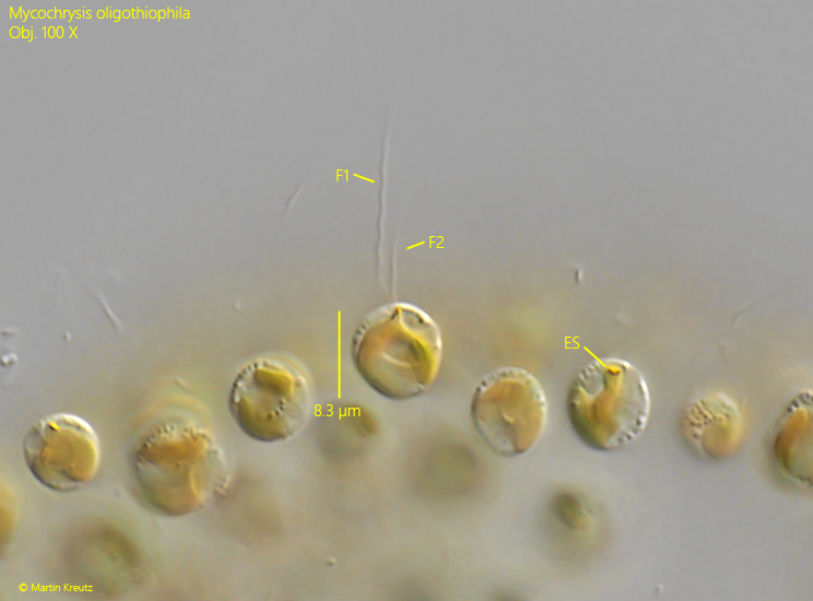
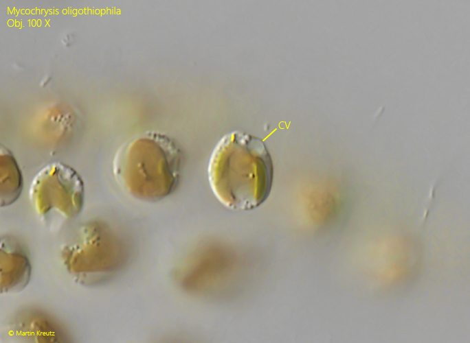
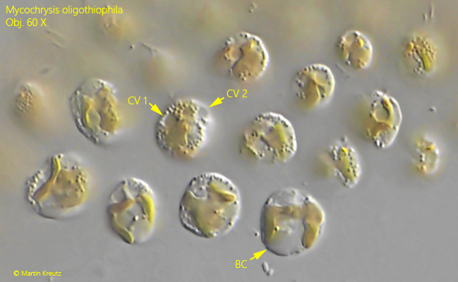
The colonies in my population consisted of about 50–100 cells. These sit at the end of the stalk in the shape of a mushroom cap. The cells are broadly ellipsoid and can only be examined in more detail with difficulty, as they are quite sensitive to the coverslip. However, I was able to clearly observe the flagella of unequal length (s. fig. 3) and the apically located contractile vacuole (s. fig. 4). Some cells possessed two contractile vacuoles, just as Skuja describes (s. fig. 5). The cells in my population were between 6–9 µm long and thus somewhat smaller than the range of 8–15 µm given by Skuja.
Fig. 1 a-b:Mycochrysis oligothiophila. L = ~250 µm (of colony). Two focal planes of an unbranched colony. The stalk is consisting of a granular and delicate gelatinous mass. Obj. 40 X.
Fig. 2 a-b:Mycochrysis oligothiophila. L = ~150 µm (of colony). Two focal planes of second unbranched colony. The shape of the stalk is visible by the adhering bacteria. Obj. 40 X.
Fig. 3:Mycochrysis oligothiophila. L = 6–8.3 µm (of cells). The two flagella (F1, F2) of the cells have an unequal length. ES = eyespot. Obj. 100 X.
Fig. 4:Mycochrysis oligothiophila. L = 8.9 µm. In this cell the apical contractile vacuole (CV) is visible. Obj. 100 X.
Fig. 5:Mycochrysis oligothiophila. Some cells in apical and lateral view. In one cell two contractile vacuoles (CV1, CV2) are visible. BC = body of chrysolaminarin. Obj. 100 X.