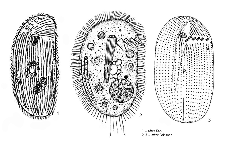
left body margin almost straigt, right margin convex
length 70–140 µm
nassulid frange fragmented, reach to middel of dorsal side
oral basked composed of 24 rods, widened in front
pellicle distinct by alveolar layer with polygonal structure
mucocysts inconspicuous (visible after staining), extrusomes absent
macronucleus spherical or slightly ellipsoid, with reticulate chromatin
micronuclus very small, close to macronucleus
contractile vacuole in body center, excretion porus ventral
some elongated caudal cilia, about 20 µm
Nassulides pictus
I found Nassulides pictus in a moss sample that came from the branch of a tree. Kahl (1935) also found his specimens in moss, while Foissner (1980) found this species in small ponds and puddles of meltwater.
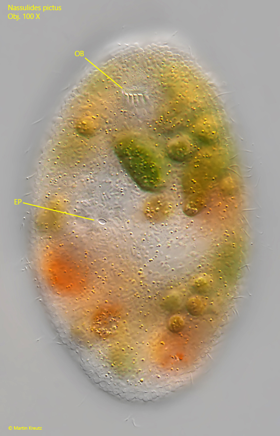
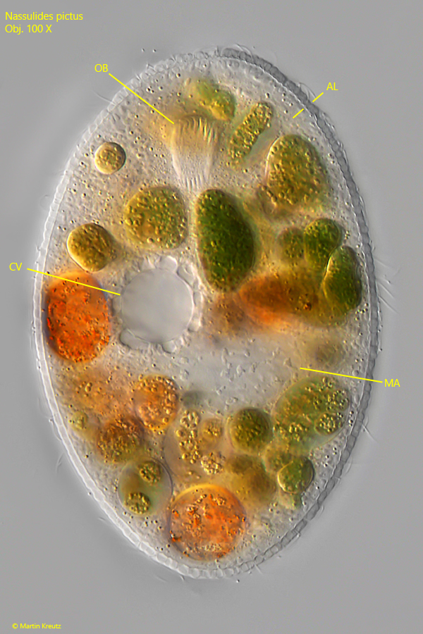
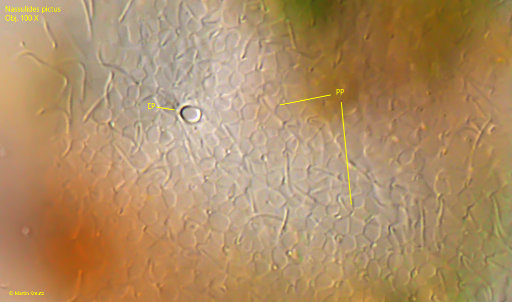
The specimens of my population were 70–90 µm long and thus corresponded approximately to the length also given by Kahl. Foissner, on the other hand, observed specimens up to 140 µm. The cytoplasm is completely filled with greenish and orange food vacuoles of phagocytized cyanobacteria. The contractile vacuole is located centrally with a ventral excretory spore. This lies below the basket, but slightly shifted to the right (s. fig. 1). My specimens had a rather ellipsoidal macronucleus, just as Kahl has drawn it (see drawing 1, above). I could not observe any caudal cilia as described and drawn by Foissner. The pellicle of my specimens were conspicuously thick due to a thick alveolar layer (s. fig. 2), which has a polygonal structure (s. fig. 5).
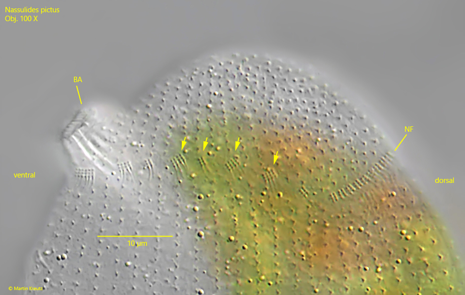
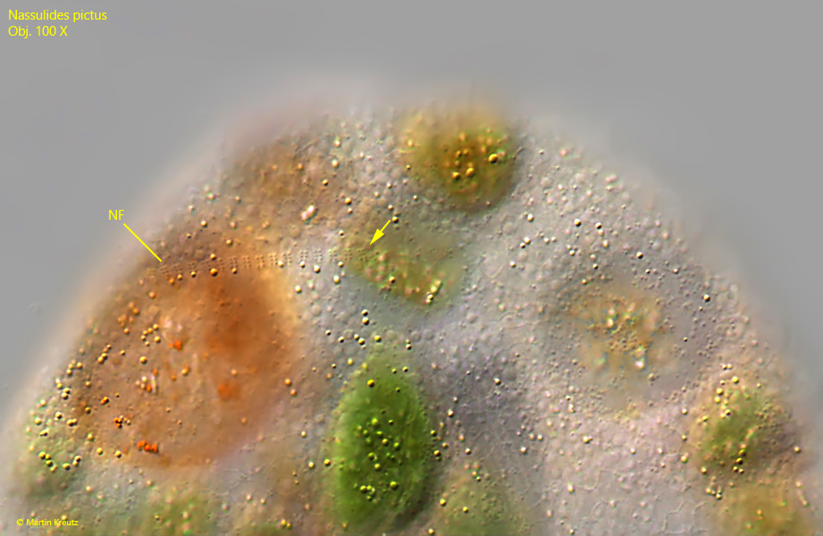
An important identification feature of nassulid ciliates is the course and shape of the hypostomial ciliary band (= synhymenium = nassulid frange). In 2002, Nassula picta was transferred to the genus Nassulides by Foissner et al. and renamed Nassulides pictus due to the fragmentation of the nassulid frange (s. fig. 3). I was able to observe this well and it corresponds to the drawings and descriptions of Foissner et al. (2002). The nassulid frange starts below the basket and runs over the left side of the body to the middle of the dorsal side (s. fig. 4).
Fig. 1:Nassulides pictus. L = 85 µm. Ventral view of a slightly squashed specimen. The focal plane is on the excretion porus (EP) in mid-body below the oral basked (OB), but slightly shifted to right. Obj. 100 X.
Fig. 2:Nassulides pictus. L = 85 µm. Focal plane on the contractile vacuole (CV) surrounded by several auxiliary vacuoles. The macronucleus (MA) of this specimen is ellipsoidal. Note the distinct alveolar layer (AL) of the pellicle. OB = oral basket. Obj. 100 X.
Fig. 3:Nassulides pictus. The nassulid frange (NF) starts below the basked (BA) and running over the left side to the middle of the dorsal side. Note the fragmentation of the nassulid frange on the left side (arrows). Obj. 100 X.
Fig. 4:Nassulides pictus. The nassulid frange (NF) ends in the middle of the dorsal side (arrow). Obj. 100 X.
Fig. 5:Nassulides pictus. Focal plane on the polygonal pattern (PP) of the alveolar layer. EP = excretion porus. Obj. 100 X.