So far, I have found only a few colonies of Nephrodiella minor in the Lauchsee Moor (Austria) in June 2024. Due to the small size of the cells, the colonies can easily be mistaken for those of bacteria or cyanobacteria at low magnifications.
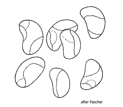
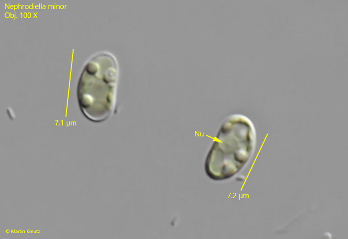
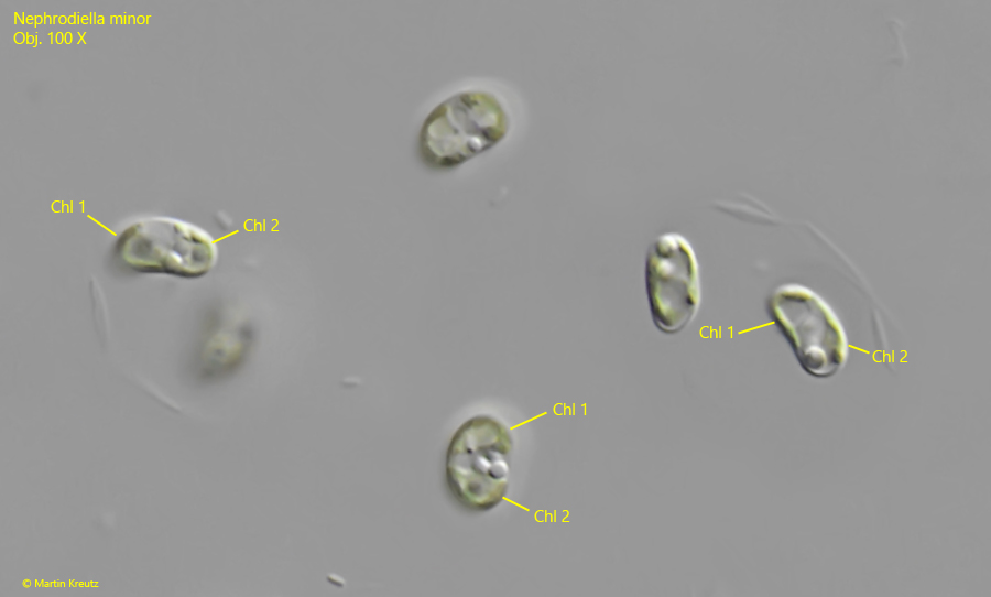
The cells of Nephrodiella minor are kidney-shaped with rounded ends. The cells appear somewhat plump in shape. The cells are embedded in a gelatinous matrix, which does not contrast under DIC. The cells may contain 1 or 2 chloroplasts, which are distinctly yellow-green. Apart from droplets of chrysolaminarin, no other components such as protein crystals are visible in the cytoplasm. The nucleus is located centrally on the concave inner side of the cells.
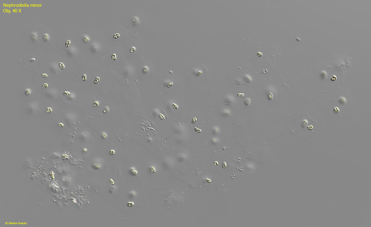
Fig. 1:Nephrodiella minor. L = 320 µm (of colony). A colony of widely spaced cells in a very delicate gelatinous mass. Obj. 40 X.
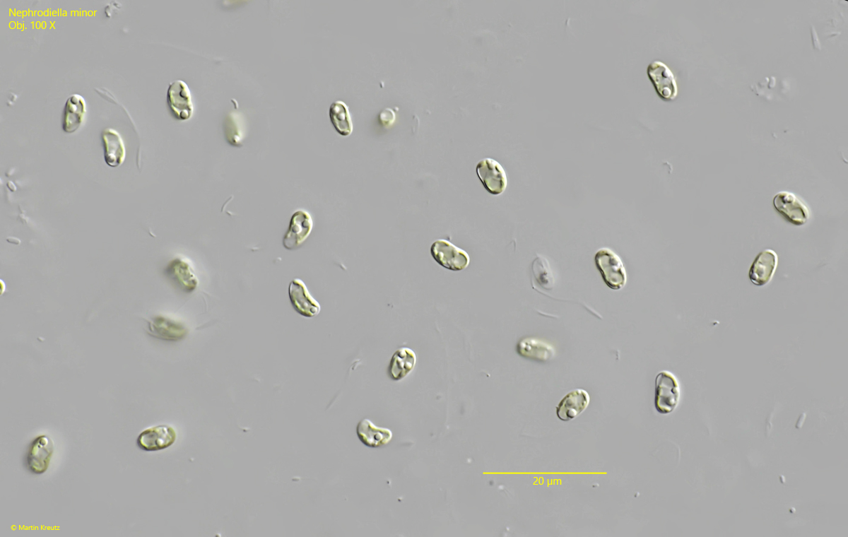
Fig. 2:Nephrodiella minor. L = 7-8 µm (of cells). Some cells in the squashed colony. Obj. 100 X.
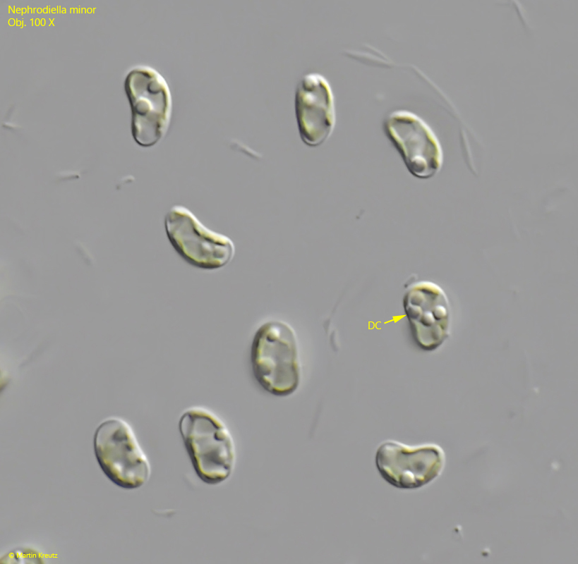
Fig. 3:Nephrodiella minor. L = 7.0–7.2 µm (of cells). Some cells in detail. Note the droplets of crysolaminarin (DC). Obj. 100 X.
Fig. 4:Nephrodiella minor. L = 7.1–7.2 µm. Two cells in a squashed colony. Nu = nucleus. Obj. 100 X.
Fig. 5:Nephrodiella minor. Some cells with two chloroplasts (Chl1, Chl 2). Obj. 100 X.
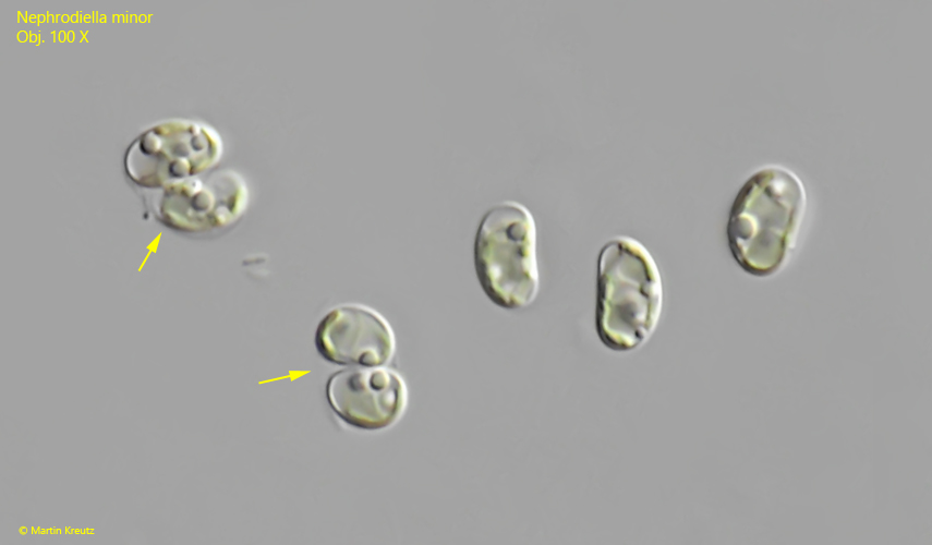
Fig. 6:Nephrodiella minor. Two cells in the process of cell division (arrows). Obj. 100 X.