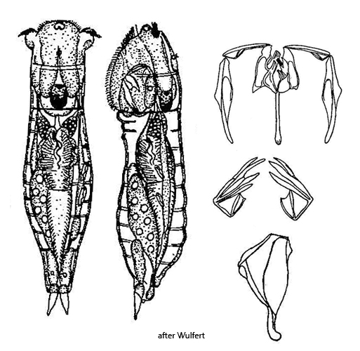
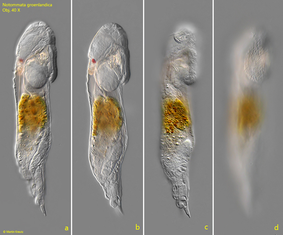
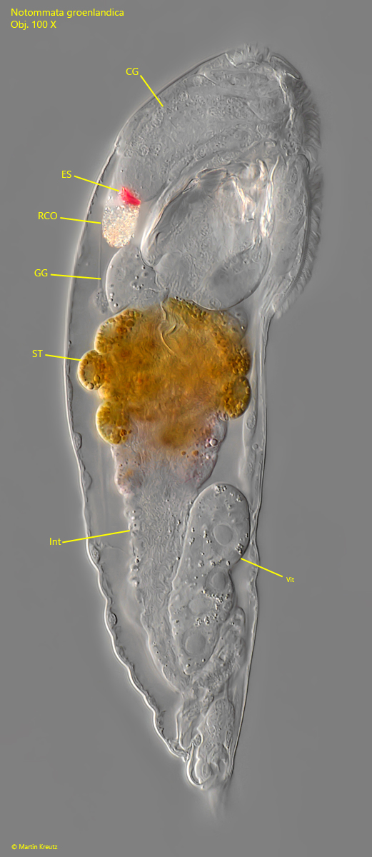
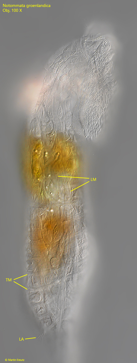
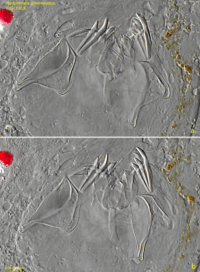
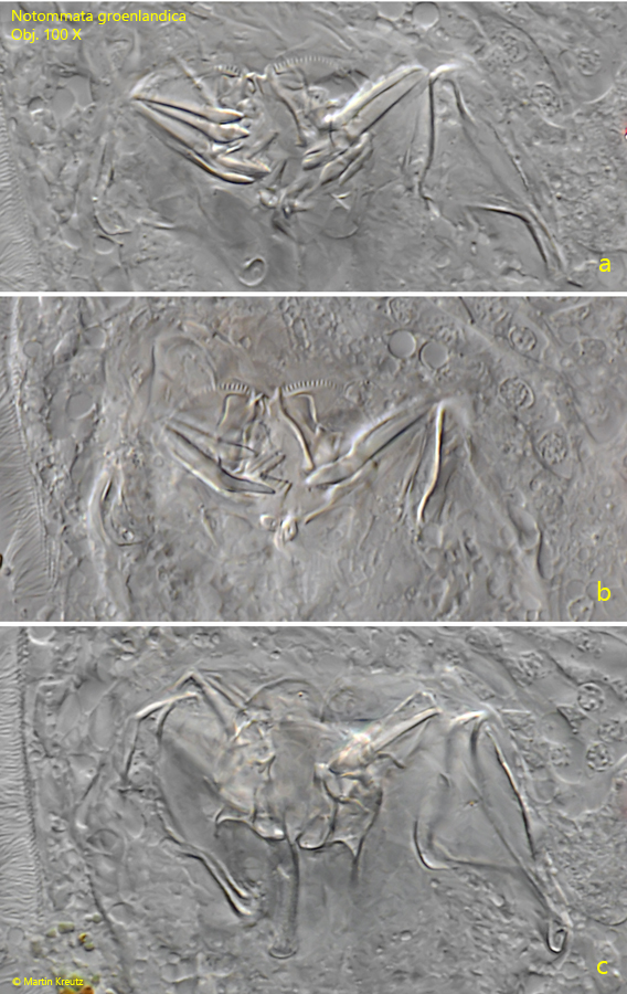
So far, I have found Notommata groenlandica in August 2025 in the Sima Moor in Austria and in June 2026 in the Schwemm Moor, also in Austria. The locations match the description by Koste (1978), who stated that the species mainly occurs in Sphagnum ponds.
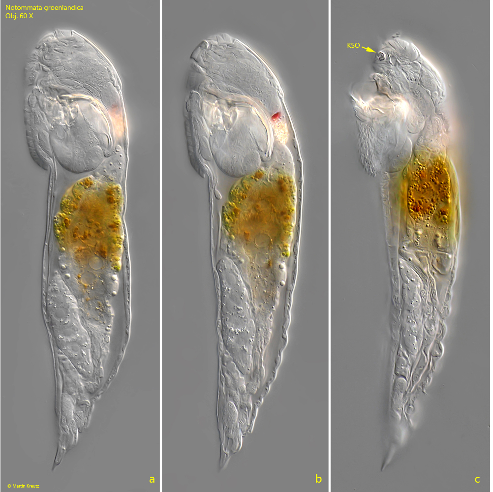
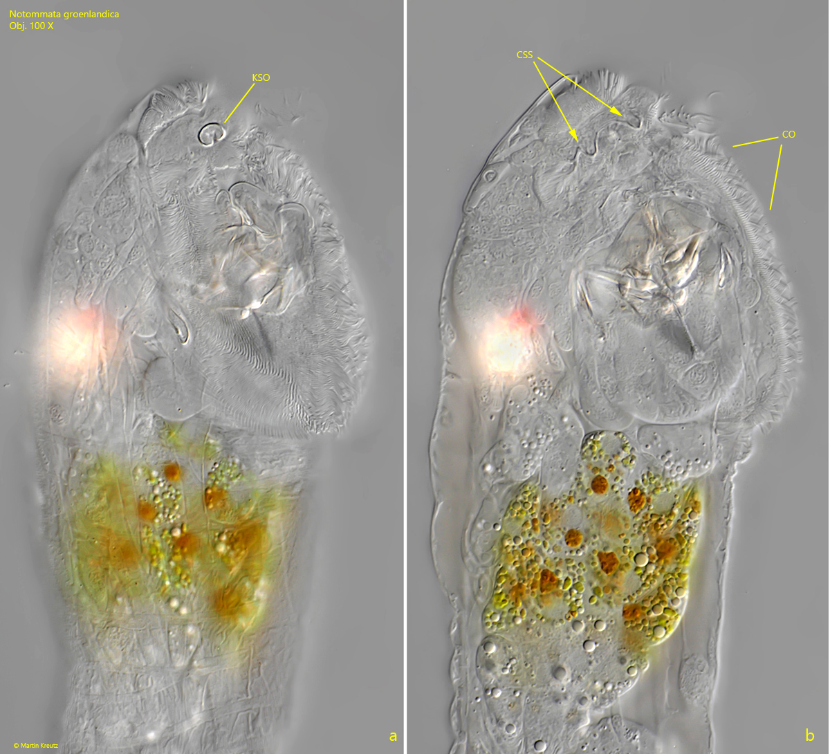
Notommata groenlandica possesses some features that allow for clear identification. On the head, below the rostrum, there is a kidney-shaped organ (s. fig. 6 a). It is best seen in ventral view in slightly squashed specimens. In lateral view, it is often not in focus. This organ is described by Koste as a “kidney-shaped crystal.” According to my observations, however, it is not a crystal, but rather a protrusion of the cuticle. If the focus is adjusted a bit deeper, a, clamp-shaped structure becomes visible (s. fig. 6 b). It appears to lie beneath the cuticle and is clearly connected to the kidney-shaped organ. Nothing is known about the function of these structures. Possibly, it is related to the feeding behavior of Notommata groenlandica, which consists of the contents of algal cells. For this, Notommata groenlandica bites a hole in the cell wall of the alga and then sucks out its contents. Algae of the genus Netrium are preferably sucked out. I myself was not able to observe this process.
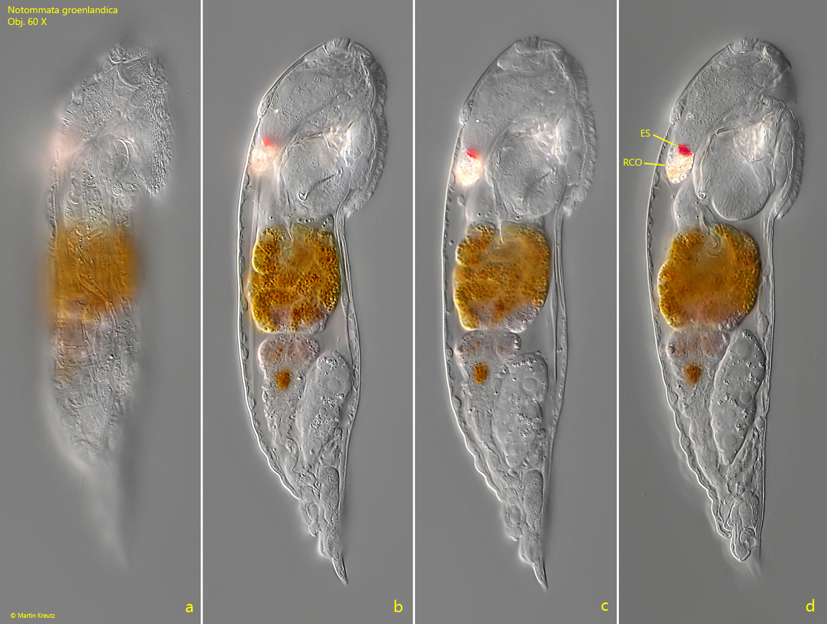
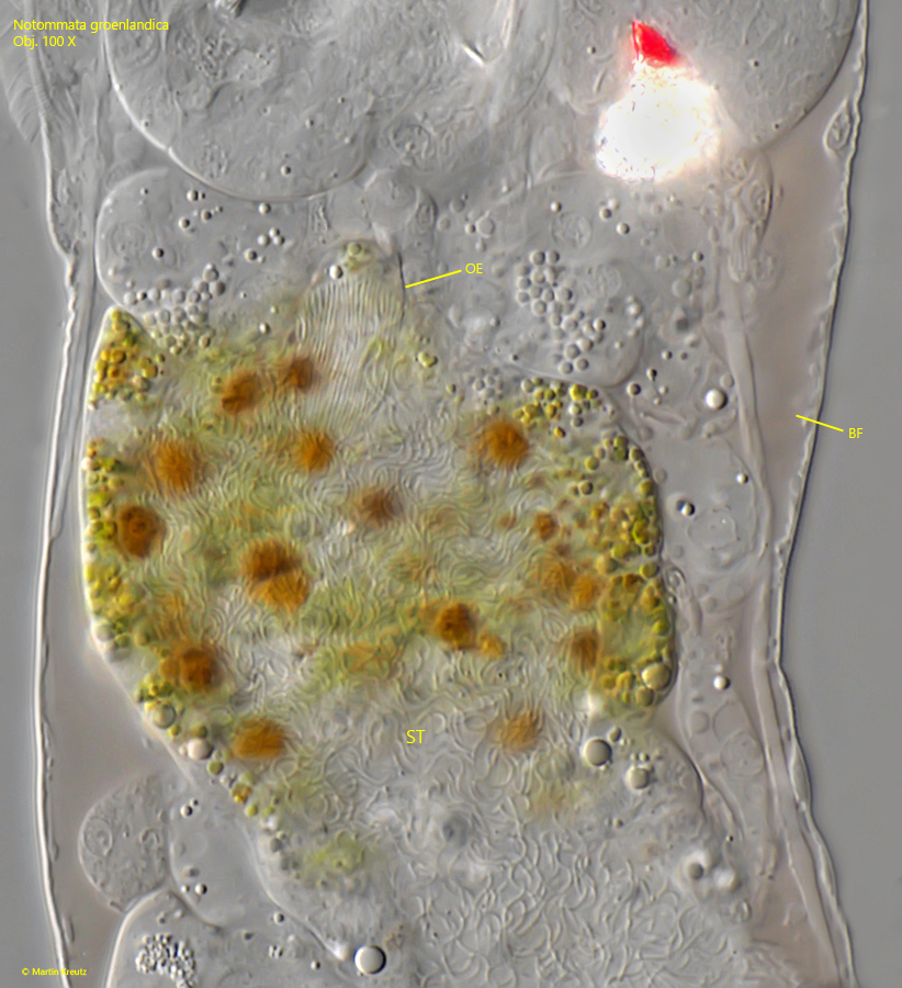
Another special feature of Notommata groenlandica is a reddish or brownish colored body fluid, which is particularly observed in older specimens (s. fig. 7). The stomach is usually distinctly yellow, green, and orange in color (s. fig. 4). At the upper end of the stomach, the esophagus ends into the stomach. There is a bundle of long, parallel cilia located there (s. fig. 7).
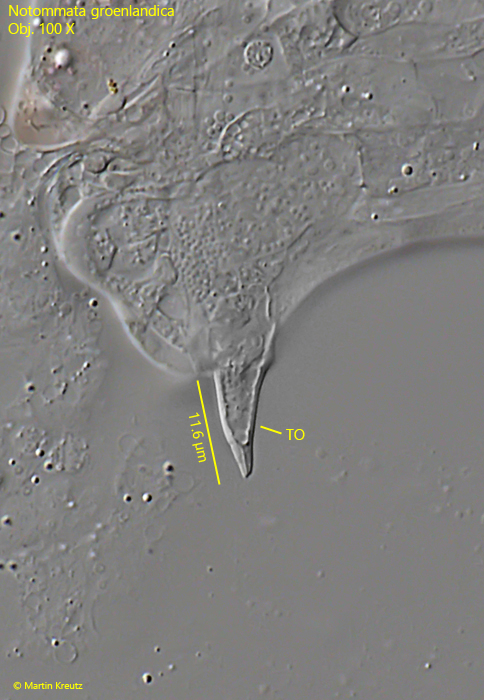
Between the conically shaped toes, there is said to be a small papilla, which I could not clearly identify, as the specimens mostly presented themselves from the lateral side under the coverslip.
Interestingly, there are differing statements regarding the presence of a bladder in Notommata groenlandica. While Voigt (1957) writes that no bladder is present, Koste (1978) mentions one. In the specimens of my population, I could not detect a bladder.
Notommata groenlandica can be confused with Notommata silpha. However, this species does not possess a kidney-shaped organ on the forehead, its body fluid is colorless, and the retrocerebral organ is small and without crystals.
More images and information on Notommata groenlandica: Michael Plewka-Freshwater life-Notommata groenlandica