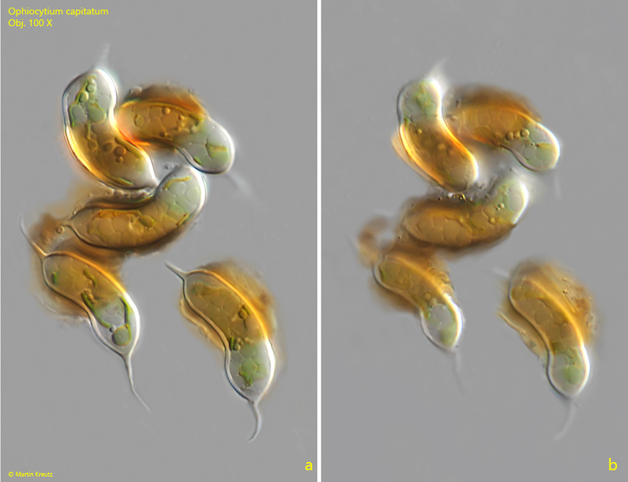
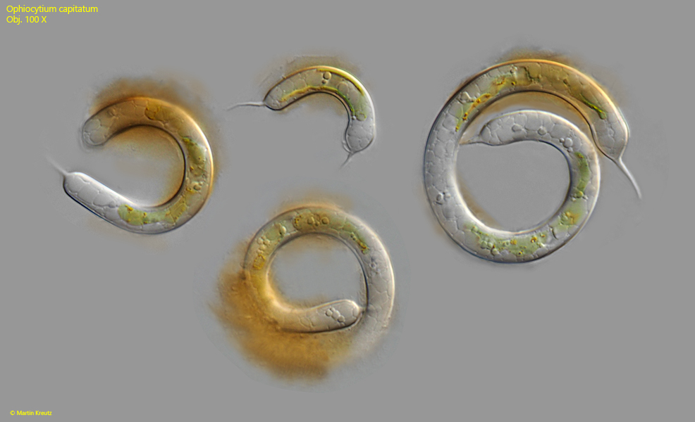
So far, I have only found Ophiocytium capitatum once in the Simmelried. In the samples, I found both solitary specimens and groups of several specimens lying close together.
The species within the genus Ophiocytium can essentially be distinguished by the following characteristics:
– number of spines – length of the cells – diameter of the cells
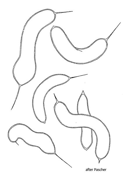
Ophiocytium capitatum has spines at both ends, which can vary in length. The cells are mostly slightly curved or almost circular and up to 150 µm long. The diameter of the cells is 5–7 µm. In my population, the diameter of the cells was 5.5–8.0 µm, which corresponds well with the data from Ettl (1977). Cells with the same shape but a diameter over 10 µm are Ophiocytium bicuspidatum.
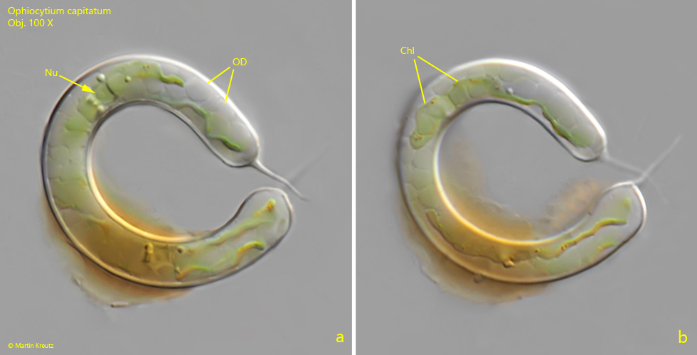
All cells of Ophiocytium capitatum in my population were partially covered with iron deposits. This phenomenon was also observed by Ettl and can occur in all species within the genus. The chloroplasts of Ophiocytium capitatum are irregularly shaped and delicately yellow-green colored. Small orange or red granules often appear in the chloroplasts, which I was also able to observe (s. fig. 3 b). The assimilation product is a colorless oil, which fills the entire cell in the form of droplets (s. fig. 3 a).
Fig. 1 a-b:Ophiocytiumcapitatum. L = 31–45 µm (with spines). Different focal planes of a collection of a few specimens, some of which are partially covered with iron deposits. Obj. 100 X.
Fig. 2:Ophiocytiumcapitatum. L = 40–145 µm (with spines). A collection of 4 specimens covered partly with iron deposits. Obj. 100 X.
Fig. 3 a-b:Ophiocytiumcapitatum. L = 99 µm (with spines). Two focal planes of a curved specimen. The cell is filled with colorless oil droplets (OD). Note the small nucleus (Nu). Chl = chloroplast. Obj. 100 X.