body slender ellipsoidal, apically rounded, posterior pointed
length 200–500 µm
oral apparatus with shape of a “6”
oral apparatus with distinct watch-glass body
distinct fringe of extrusomes forms a cortical layer
extrusomes rod-shaped, length about 6.5 µm
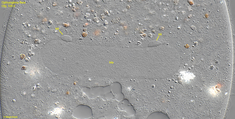
macronucleus elongated ellipsoid
one micronucleus adjacent to macronucleus
two contractile vacuoles with auxiliary vacuoles and collecting canals
each contractile vacuole with 3–4 excretion pores
Ophryoglena flava
I find Ophryoglena flava regularly, but always only sporadically and in single specimens. They are usually found in floating and decomposing plant masses. I found most of the specimens in the Simmelried.
The body shape of Ophryoglena flava is very variable. Often the specimens are completely opaque due to masses of food vacuoles and are almost round in shape. Only rarely are starving specimens found, which then have a slender shape with a slightly pointed posterior end (s. fig. 1 a-d). Such specimens are then yellow-brownish in color. The specimens swim very fast and move hectically forwards and backwards. The body is also extremely metabolic.
Like all other species of the genus, Ophryonglena flava has an oral apparatus in the shape of a “6”. The oral apparatus has a complex structure and contains the so-called watch-glass body, which is also known as Lieberkuehn’s organelle (s. figs. 3 and 4). The watch-glass body of Ophryoglena flava is clearly recognizable. It is slightly yellowish in color and its surface is finely striated (s. fig. 5 a-b). Its function is unclear. However, it is assumed that it is connected with the widening of the mouth opening when swallowing large prey.
Ophryoglena flava can be easily distinguished from other species of the genus by the two contractile vacuoles located on the right side of the body (s. fig. 2 a). They have many auxiliary vacuoles, which taper into thin collecting canals that extend very far into the cytoplasm (s. fig. 6). Each contractile vacuole has 3-4 excretory pores, which can be clearly seen when focusing on the pellicle (s. fig. 7).
Fig. 1 a-d:Ophryoglena flava. L = 190 µm. A freely swimming specimen from ventral (a, b and d) and right (c). Note the slightly pointed posterior end. Obj. 40 X.
Fig. 2 a-c:Ophryoglena flava. L = 220 µm. A slightly squashed specimen from ventral (a, b) and from left (c). Note the two contractile vacuoles (Cv 1, CV 2) located at the right side. Ma = macronucleus, Mi = micronucleus. Obj. 40 X.
Fig. 3:Ophryoglena flava. The complex oral apparatus in lateral view. Note the watch-glass body (WGB) and the endoral membrane (EM) in the deep vestibulum. Obj. 100 X.
Fig. 4:Ophryoglena flava. Focal plane on the watch-glass body (WGB) in oblique lateral view. Obj. 100 X.
Fig. 5 a-b:Ophryoglena flava. Two focal planes of the watch glass-body in a strongly squashed specimen. Note the fine striation on the surface of the watch-glass body (arrow). Obj. 100 X.
Fig. 6:Ophryoglena flava. One of the two contractile vacuoles (CV) surrounded by auxiliary vacuoles (AC) and collecting canals (CC). Obj. 100 X.
Fig. 7:Ophryoglena flava. Each contractile vacuole has 3–4 excretion pores (arrows). Obj. 100 X.
Fig. 8:Ophryoglena flava. One of the two contractile vacuoles (CV) in lateral view. Note the two cup-shaped excretion pores (EP) in lateral view. Obj. 100 X.
Fig. 9:Ophryoglena flava. L = 190 µm. The rod-shaped extrusomes (EX) are about 6.5 µm long. Obj. 100 X.
Fig. 10:Ophryoglena flava. The elongated macronucleus (Ma) with two adjacent micronuclei (Mi). Obj. 100 X.