body asymmetrically oval or broadly ellipsoid, flexible and deformable
laterally flattened
right side ciliated
left side naked, margin is curved upwards
a distinct indentation near anterior end on left side
mouth slit ventral in anterior third (only visible during ingestion of food)
length 80–180 µm, width 35–55 µm
whole margin of the body flattened with a fringe of delicate, long extrusomes
cytoplasm opaque by fine granula
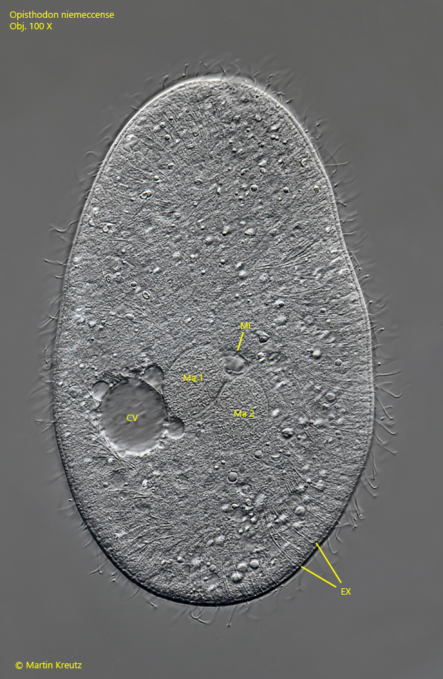
one micronucleus in the vicinity of two macronuclei
contractile vacuole near midbody, shifted dorsally
Opisthodon niemeccense
I find Opisthodon niemeccense regularly, but never frequently in my sampling sites with a sapropelic zone. Even at low magnifications Opisthodon niemeccense can be recognized by its mostly oval shape with a transparent margin. In brightfield illumination the ciliate appears brownish or even black due to the dense, fine granules in the cytoplasm. In DIC the ciliate appears bright and opaque.
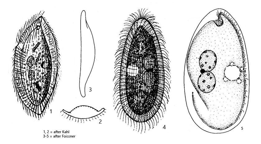
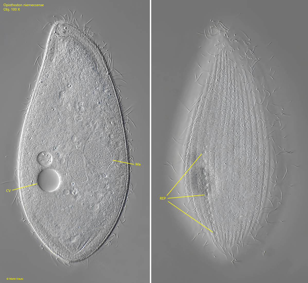
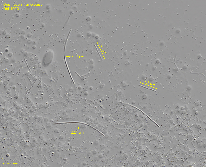
Opisthodon niemeccense has a characteristiv cavity at the anterior end on the left (unciliated) side of the body. Unfortunately, I have not yet been able to document this because all the specimens I have photographed faced with their right (ciliated) side to the coversplip. In the specimens of my population I found the contractile vacuole below the cell equator (s. figs. 1 a, 2 a and 3 a), what is in contrast to the descriptions and drawings by Kahl and Foissner (s. drawings above). In addition, I was able to document a row of 10–15 excretory pores on the right side above the contractile vacuole (s. fig. 3 b), which to my knowledge has not been described before. In addition, I investigated the extrusomes in Opisthodon niemeccense in squashed specimens. I was able to recognize long, curved extrusomes with a length of 22-24 µm but also a second type of shorter, straight rods with a constant length of 6.7 µm.
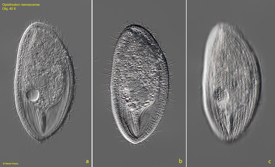
Fig. 1 a-c:Opisthodon niemeccense. L = 124 µm. Three focal planes from right of a freely swimming specimen. Obj. 40 X.
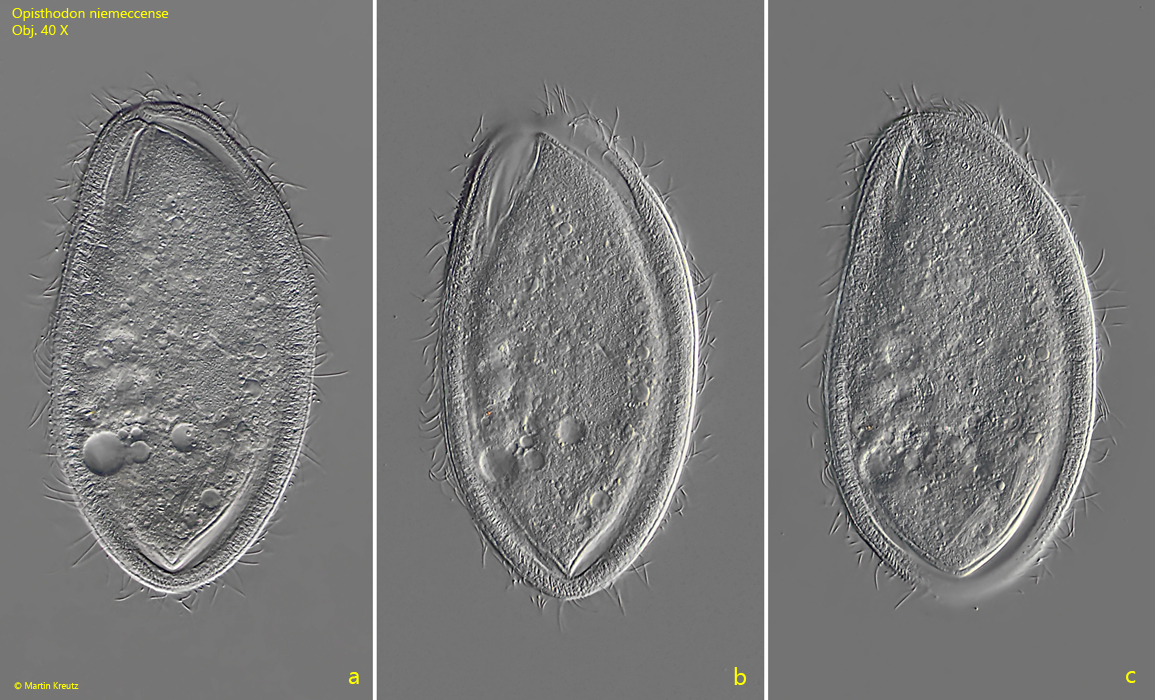
Fig. 2 a-c:Opisthodon niemeccense. L = 145 µm. A second, freely swimming specimen from right. Obj. 40 X
Fig. 3 a-b:Opisthodon niemeccense. L = 130 µm. Two focal planes of a slightly squashed specimen from right. Note the row of excretion pori (REP) of the contractile vacuole (CV). Ma = macronucleus. Obj. 100 X.
Fig. 4:Opisthodon niemeccense. The micronucleus (Mi) between two oval macronuclei (Ma 1, Ma 2) in a squashed specimen from right. Note the fringe of long, curved extrusomes (EX). CV = contractile vacuole. Obj. 100 X.
Fig. 5:Opisthodon niemeccense. In a strongly squashed specimen curved extrusomes with a length of 22–24 µm are visible as well as straight rods with a length of constantly 6.7 µm. Obj. 100 X.